Download

1 / 22
230 likes | 405 Vues
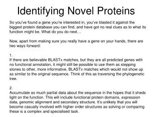
Novel labeling technologies on proteins. a). h n. h n. Protein 1. BFP. FRET. +. Fluorescence. Fluorescence. GFP. Protein 2. b). 433 nm. 433 nm. CFP. PKA + ATP. pS. 14-3-3 t. 476 nm. FRET. phosphatase. Substrate peptide. 527 nm. YFP. c). Protein or peptide. GFP. N. N.

E N D
a) hn hn Protein 1 BFP FRET + Fluorescence Fluorescence GFP Protein 2 b) 433 nm 433 nm CFP PKA + ATP pS 14-3-3 t 476 nm FRET phosphatase Substrate peptide 527 nm YFP c) Protein or peptide GFP N N C C • Fig.1 Fluorescent protein labeling (Gene fusion approaches) • Protein-protein interaction is detected by FRET using two different GFP analogues • Detection of kinase activity using GFP-fusion protein • Application of domain insertion for functional switching
a) Translated protein Puromycin-fluorophore conjugate P site A site mRNA without stop codon Aminoacyl-tRNA Ribosome C-terminus labeled protein b) Florpuro Cy5-puro
c) α-helical CCxCC domain S fluorescent S S S d) protein Ni2+ Oligo-histidine tag FLASH Non-fluorescent
a) R R DNA 1) DMAP HS- QD 2) H2O R R R R R= R= DNA DTT H2N- QD Surface modification of quantum dots Making of Barcode by encapuslating Q-dots in polymer beads
(a) Cell lysate (protein mixture) Each protein is detected by direct labeling, labeled antibody, mass spectrometry or SPR Ligand array (b) Protein or other molecule, that is interested Molecular interaction is detected by direct labeling, labeled antibody, mass spectrometry or SPR Fluorescent label Protein array
a) Sample protein Protein (covalently immobilized) Various proteins are spotted on a membrane Protein (adsorption) PEG a) Immobilization using SAM d) c) Immobilized protein Polyacrylamide gel pad NH O=C NH N O = CHO C OH Glass plate Glass plate Protein immobilization in gel pad BSA Protein is immobilized covalently on Glass slide b)
(a) (b) Target protein Protein complex Protein complex Ligand (Antibody) Ligand (Antibody)
MALDI TOF MS Matrix assisted laser desorption ionization – Time of flight Mass spectroscopy α-cyano-4-hydroxycinnamic acid CHCA
MS fingerprint Peptide fragments Detecting MS profile of fragments Protein Electrophoresis gel MS analysis Trypsin digestion comparison Database of protein sequences Database of predicted MS fingerprints of each known proteins Simulation of trypsin digesting pattern
Peptide MS fingerprint and Peptide sequence Tag Enzymatic labeling of stable isotope coding of proteomics Proteins from two distinct proteome are digested with protease in normal water or isotopically labeled water. Isotobe code is labeled in every C-terminus of the digested peptides. Then, two samples are combined and analyzed by LC-MS/MS. Expression level of proteins between two states can be estimated. Amino acid sequence of selected peptide fragment can be identified, too. fmollevel is needed to keep practical sped
Quantified Proteome(Labeling of stable iostope) SILAC (Stable Isotope Labeling by Amino acids in Cell Culture) Harvest cells Combine and cell lysis Proteolysis after denaturation and reduction d3Leu, d3Met, d2Tyr) , d3Ser, 13C6Arg, 13C6Lys Can’t be applied to animals mLC-MS in vivo stable isotope labeling of proteome sample Cells are grown in normal media or isotopically labeled media. Mass tags are incorporated into every protein. An equivalent number of cells for each sample are combined and processed for MS.
Trasfection to cell sample Immuno-precipitation General strategy for investigating intracellular protein interaction with MS analysis. Cell lysis MS analysis b) Identification of interacting regions in protein-protein interaction After immuno-precipitation, the complex is crosslinked with this cross-linker, then was digested with protease to make fragment couple linked with the reagent. Stable isotope coded cross-linker
Isotope-coded Affinity Tag (ICAT)法 Isotope-coded Affinity Tag (ICAT) Comparison of protein expression levels between two samples by using fragments containing cysteine residue Purified with Avidin column
a) P Biotin Biotin b) base H2O m/z=446 fragment Application of ICAT to phosphoproteome a) Scheme of isolating phosphorylated peptide b) Reaction scheme of the chemical conversion of phosphoserine residue to a biotinylated moiety.