Download
1 / 48
490 likes | 947 Vues
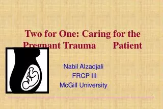
Two for One: Caring for the Pregnant Trauma Patient. Nabil Alzadjali FRCP III McGill University. CASE 1 25 Yrs F, 35 wks Preg. PC : MVC PMH : nil, Rh +ve, HPI : Driver, belted, rear ended by another car, air bag deployed Complaining of occasional abdominal pain,

E N D
Two for One: Caring for the Pregnant Trauma Patient Nabil Alzadjali FRCP III McGill University
CASE 1 25 Yrs F, 35 wks Preg. PC : MVC PMH : nil, Rh +ve, HPI : Driver, belted, rear ended by another car, air bag deployed Complaining of occasional abdominal pain, ?cramping. Unsure about fetal movements. Very concerned regarding fetal well being. ABC stable. BP 120/70 HR 88 RR 15 No signs of injuries on exam. FHR 140, No uterine contractions palpable. No guarding. No lap belt sign. No PV bleeding. Os Closed • How do we manage this patient?
CASE 2 20 Yrs F, 30 weeks gestation Struck by truck across the street from hospital. Cardiac arrest at scene. U/G Technician have intubated and started CPR. Down time about 5 minutes. Arrival in ER, Pulseless Electrical Activity. • How do we manage this patient?
Incidence • Physiological Alterations • Anatomical Alterations • Unique Problems in the Gravid Abdomen • Prehospital Considerations • Diagnostic Studies • Management of trauma • Unstable Mother • Stable Mother • Perimortem Cesarean Section
Incidence • The Leading cause of non-obst. mortality - 46% • Trauma during pregnancy - 7% • Causes of Trauma (1) • MVA 54.6 % • Domestic abuse & Assault 22.3 % • Falls 21.8 % • Penetrating inj. 1.3 % • < 1% of trauma admissions are pregnant • Preterm Labor in 11.4 % & P. Abruption in 1.58 % (1)Connolly A, Katz VL, Bash KL, et al: Trauma and pregnancy. Am J Perinatol 14:331-336, 1997
Supine Hypotensive Syndrome(1) (1) Milson I, Forssman L: Factors influencing aortocaval compressionin late pregnancy, Am J Obtst Gynecol 148: 764-771, 1984
Respiratory • Respiratory alkalosis • Reduce oxygen reserve (reduced FRC 20% & increased O2 consumption by 15 %) • Residual volume decreased by 40% • Respiratory rate increased • Impaired buffering capacity
GI • Intestine are concentrated in upper abdomen • Decrease GI motility • Decrease peritoneal irritation • GU • Bladder is displaced upward >10 wks • Dilitation of renal pelvis and ureters
Alterations in Anatomy • 1st trimester uterus is thick walled and intra-pelvic • Out of pelvis > 12 wks. • Second trimester uterus contains large amount of amniotic fluid • Third trimester uterus is thin walled, large Fetal head engaging pelvis • At 36 weeks uterus reaches costal margin
Injuries unique to pregnancy • Premature Contractions • Rarely progress to preterm delivery • Tocolysis is not proven in trauma.(1) • Abruptio Placentae • Different elastic properties in uterus & placenta “shearing” • 3 % of minor trauma and upto 50 % in severe trauma (1) GoodwinTM, Breen MT: Pregnancy outcome and fetomaternal hemorrhage after noncatastrophic trauma, Am J Obstet Gynecol162: 665-671, 1990.
Uterine Rupture • Rare, 0.6 % of severe abdominal trauma (1) • Direct trauma after 12 wks of gestation • Prior Surgery (C/S or Myomec.) the risk • Maternal-Fetal Hemorrhage • Trimesters 1 3%, T2 12%, T3 45% • 4-5 X more common in injured pregnant women • Causes isoimmunization & fetal death • Kleihauer-Betke test - volume of fetal blood • .01- .03 cc sensitize, 5 cc +ve KB Test. • To determine amount of Rhogam needed 1. Pearlman MD, Tintinalli JE, Lorenz RP: Blunt trauma during pregnancy, N Engl J Med 323:1609, 1990
Special Considerations • Blunt Abdominal Trauma • Penetrating Abdominal Trauma • Stabbing injury • Gunshot injury
Blunt Trauma • Injuries • Head injury most common • Retroperitoneal hemorrhage • Abruptio placenta • DIC • Uterine Rupture • Seatbelts – 3 Points Restraints • 1/3 – ½ improperly or don’t use belts • Unbelted is at 2.3X to give birth <48 hrs & 4.1X fetal death
Penetrating Injury GSW’s • Gravid uterus alter injury pattern to the mother. • If missile enter upper abdomen; increased probability of harm (upto 100%). • If enters below uterine fundus visceral injury less likely (0%) • Awwad et al (1) • Fetal death rate is 67% • 38 % for injuries above the uterus. (1) Awwad JT et al: High-velocity penetrating wounds of the gravid uterus: Review of 16 years of civil war, Obstet Gynecol 83:259, 1994.
Stabbing Injury • Rare rare, only 19 cases reported in literature • Morbidity 93 % - Mortality 50 % • Many advocate exploratory laprotomy since uterus laceration is devastating b/c of its enlarged circulation. • Meizner et al (1) • An injury to uterus can rapidly change to a hypotensive emergency. • It is difficult to know the size and depth of uterine rupture (1) Meizner I, Potashnik G: Sharpnel penetration in pregnanc resulting in fetal death, Isr J Med Sci 24:431, 1988.
Pre-hospital Consideration • Oxygen • Shock should be anticipated • ED should be notified early, GA >24 wks • Transport in L lateral position (GA > 20 wks) National Association of EM Physician, 1997 “PASG – class III intervention” worsen the supine hypotension
Modalities for Evaluating Trauma • Plain Films – X-rays • Ultrasound • CT & MRI • Cardiotocographic Monitoring • DPL • Laparotomy
Plain Films • Risk of 1 rad to fetus is approx. 0.003 • < 5-10 rads causes • No risk on congenital malformation, abortions or intra-uterine growth ret. • Smaller risk of increase in childhood cancer • Radiation doses > 10 rads • 6 % chance of severe mental ret. • < 3 % chance childhood cancer.
Rosenstein M:Handbook of selected organ doses for projections common in diagnostic radiology. HEW publication(FDA) 89-8031. Rockville, MD. US Dept. Of Health And Human Services, Centre For Devices And Radiologic Health, 1988.
Ultrasound • Best modality to assess both fetus and mother • Not sensitive: • Colonic lesions • Biliary tree lesions • Sub-placental hematoma • Safe procedure
CAT SCAN • Complementary to U/S & DPL • Penetrating wounds of flank & back • Can miss diaphragmatic and bowel injuries • Portability • Spiral CT reduces radiation exposure by 14-30 %
Rosenstein M:Handbook of selected organ doses for projections common in diagnostic radiology. HEW publication(FDA) 89-8031. Rockville MD,. US Dept. Of Health And Human Services, Centre For Devices And Radiologic Health, 1988.
Cardiotocographic Monitoring • FHR • Rate (120-160) • Beat-to-beat variability • Baseline variability • Decelerations, esp. late
Cardiotocographic Monitoring Variability:
Cardiotocographic Monitoring Decelerations: Early and Late
Cardiotocographic Monitoring Decelerations: Variable
Diagnostic Peritoneal Lavage • CT & U/S are better in stable patient. • Hypotensive unstable pt and if bedside U/S is not available • Can be performed in any trimester • Gravid uterus does not reduce the accuracy of DPL for OR • Limited in detecting bowel perforation and does not assess retroperitoneal hemorrhage or intra-uterine pathology
Diagnostic Peritoneal Lavage • Rothenberger et al (1) • n=12 (4 Supra umbilical & 8 infra umbilical) • Sensitivity 100 % (8 internal bleeding confirmed by lapratomy), • Specificity 100 % ( 4 no bleeding) • No Complications from the procedure • Esposito et al (2) • n=40 , 13 had DPL • PPV = 100 % • Rothenberger DA, et al:Diagnostic peritoneal lavage for blunt trauma in pregnant women, Am J Obstet Gyneco 129:479-48,1977. • Eposito TJ, et al: Evaluation of blunt abdominal trauma occurring during pregnancy, J Trauma 29:1628-1632, 1989.
Management • Avoid distractions and avoid focus on the fetus • Be aggressive! But temper with common sense. • An apparently stable mother may be compensating at expense of the fetus • If < 24 weeks, intermittent fetal doppler • If > 24 weeks, then continuous cardiotocographic monitoring to assess FHR and uterine activity
I. Initial maternal Resuscitation Airway Assess & control Preoxygenate and sellick’s maneuver is important before intubation Breathing Assess and manage Place CT in 4th intercostal space Circulation Assess maternal circulation IV access Telt to left if > 20 wks
Management • The hemodynamically unstable mother • The hemodynamically stable mother
Stable fetus Minor trauma does not exclude significant fetal injury; 1-3 % of all minor trauma results in fetal loss from placenta abruption. (1) Asymptomatic mother or with no obvious abdominal injury needs monitoring for feto-placental pathology (1) Pearlman MD, Philip ME: Safety belt use during pregnancy, obstet Gynecol 88: 1026, 1996 III. The hemodynamically stable mother
1. Pearlman MD, Tintinalli JE, Lorenz RP: Blunt trauma during pregnancy, N Engl J Med 323:1609, 1990 Pearlman et al (1) • Minimum 4 hrs CTG monitoring • Extended to 24 hrs if : . >3 contractions per hour . Persistent uterine tenderness . Non reassuring fetal monitor strip . Vaginal bleeding . ROM . Serious maternal injury present • All placental abruption were detected within 4 hrs • 70 % of pt required admission. • All discharged home subsequently had live birth.
III. The hemodynamically stable mother Unstable fetus • Fetal death rates are 3-9 times higher than mat. • No infant survive if there is no fetal heart tone before C/S • Morris et al(1) • Heart tone is best survival marker for f. to undergo C/S • If fetal heart tone is present and the GA is > 26 wks the survival is 75% • 60 % of fetal death occurs with under use of CTG and delay recognition of fetal distress.
Perimortem Cesarean Section • ~200 successful cases reported in the literature • Maternal CPR <5 minutes, fetal survival excellent • <23 weeks gestation survival chance is 0% • Maternal CPR >20 minutes, fetal survival unlikely
Perimortem Cesarean Section • 4 Minute Rule: Maternal CPR for 4minutes, Infant should be delivered by the 5th minute.
Perimortem Cesarean Section • Technique: • Make sure it is indicated first and that resuscitative team is ready • Vertical incision from xyphoid to pubis • Continue straight down through abdominal wall and peritoneum • Cut through uterus and placenta (if anterior) • Bluntly open uterus and remove fetus • Cut and clamp cord
Summary • Anatomic and physiologic changes • Vigorous fluid and blood replacement • Treat the mother first and treat her just like any other trauma patient • High index of suspicion for blunt or penetrating uterine trauma & abruptio placenta. • Consider perimortem C/S in unstable women or cardiac arrest with viable fetus after 24 wks.
When to Intervene and Consult • EARLY !
Remember What is Best for the Mother is Best for the Fetus!