Download
1 / 16
380 likes | 1.85k Vues
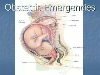
Obstetric Ultrasound. By . Alaa malki. 1- Is usually obtained twice during the normal pregnancy: (18-24 weeks) , ( 32-38 weeks ). 2- It can be done before 18 weeks: -to confirm intra-uterine pregnancy . -estimation of G.A.

E N D
Obstetric Ultrasound By .Alaamalki
1- Is usually obtained twice during the normal pregnancy: (18-24 weeks) , (32-38 weeks). 2- It can be done before 18 weeks: • -to confirm intra-uterine pregnancy . • -estimation of G.A. • -exclude abnormal pregnancy (molar or ectopic ). • -multiple pregnancy .
QUSTION ALWAYS SHOULD BE ASKED TO THE PREGNANCY WOMEN BEFROE SCANNING: - LMP. - any cardiac problems. - any previous abnormal baby . - bleeding? - any abnormal pain ? - medication ?? - diabetic ?? SCANNING : -Transvaginal: for early pregnancy on the first 9 wk -Transabdomenal.
GESTATIONAL SAC : -its usually visualized from 31 days (4+3 wks) =(2-3 mm)or (5+3 wks). -it will appear as circle area surrounding by thick bright ring at fundal area. -it grow approx. (1mm/day) -at (8 weeks) amniotic cavity expands rapidly . -at the end of the first trimester, the amniotic and chorionic membrane becomes fused .
YOLK SAC: -appear as circle mass with the G.S. -it can be identified at (35 days). -it grow slowly until reach a maximum of (6 mm)at (10 wks). -the identification of the yolk sac will be very difficult after (12 wks).
EMBRYO: -(4-9) wks(embryo). -(10-40) wks(fetus ). -it can be visualized from (5 wks) ,on this stage CROWN-RUMP-LENGTH (CRL) can be measured. -the embryo grow (1 mm/day ) -cardiac activity (heart rate ) seen (6-9 wks) -about ( 9 wks) placenta can be seen -at ( 8 wks ) upper + lower lumb can be seen more clearly .
NUCHAL TRANSLUCENCY (NT): Seen between ( 11-14 ) weeks.
Second trimester -it called ( routine anomaly scan ) (18-24 wks ). -optimal time to examine fetal heart at (23-24 wks). -fetal anatomy ,placenta ,amount of amniotic volume ,fetal movement ,measurements ,ovary ,uterus , both adnexa .
1- Biparital diameter (BPD) 2- Head circumference (HC)
5- FETAL POSITION:(cephalic-breach) 6- PLACENA SITE