
Obatoclax Biodistribution in MLL Leukemia NOG Mouse Model
This study predicts Obatoclax tissue penetration in NOG mice with MLL leukemia using modeling and simulation. The research shows high drug distribution at crucial sites, impacting leukemia treatment efficacy. The biodistribution of Obatoclax indicates significant accumulation in spleen, liver, and brain, key target areas for anti-leukemia actions. Data suggests sustained drug levels in these organs, with potential CNS penetration and liver accumulation over time. Results support pre-treatment strategies for enhancing chemotherapy in MLL+ leukemia. Simulations reveal distinctive drug exposure profiles between mouse strains, highlighting the impact of disease burden on drug elimination.

Obatoclax Biodistribution in MLL Leukemia NOG Mouse Model
E N D
Presentation Transcript
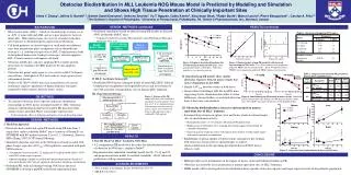
Obatoclax Biodistribution in MLL Leukemia NOG Mouse Model is Predicted by Modeling and Simulation and Shows High Tissue Penetration at Clinically Important Sites Mice injected with 1.3 million leukemia cells 3 weeks post-injection Alena Y. Zhang1, Jeffrey S. Barrett1,2, Gwenn Danet-Desoyers2, Anthony Secreto2, Vu T. Nguyen, Cathy Keefer2, Xiaochuan Shan, 2 Ralph Bunte2, Manon Lavoie3, Pierre Beauparlant3 , Carolyn A. Felix1,21The Children’s Hospital of Philadelphia, 2University of Pennsylvania, Philadelphia, PA. 3Gemin X Pharmaceuticals, Inc., Montreal, Canada Diseased Mice n = 42 1.2 mg/kg n = 21 4.8 mg/kg n = 21 PK sampling at 0.08, 0.25, 1, 2, 4, 8, 24 hr (3 mice per time point per dose level) DESIGN / METHODS (continued) RESULTS (continued) BACKGROUND Tissue sampling • MLL translocations (MLL+) , which are chemotherapy resistant, occur in ~80% of infant ALL and AML and are a poor prognostic factor in infant ALL. MLL translocations also occur in secondary leukemias after exposure to chemotherapeutic topoisomerase II poisons. • Cell death pathways are desired targets of small molecule inhibitors since their deregulation plays an important role in chemotherapy resistance; e.g. imbalanced expression of BCL-2 family proteins leads to deregulated homeostatic binding between pro- and anti-apoptotic BCL-2 family members and impairs cell death. • Obatoclax inhibits pro- and anti- apoptotic BCL-2 family protein interactions by binding to the BH3 pocket of the anti-apoptotic proteins. • Obatoclax haspotent single agent in vitro activity in MLL+ leukemias and cell lines. Adult phase I CLL trials indicate single agent activity with minimal toxicity. • NOD-scid-IL-2Rnull(NOG) mice lack natural killer cell activity, resulting in superior engraftment of human leukemia xenografts compared to other immuno-deficient mouse strains. • Performed simulations based on obatoclax pop-PK models in diseased NOG and healthy Balb/C mice • Compared simulated secondary PK parameters using WinNonLin (Pharsight, Mountain View, CA) IC90 IC50 PBPK model (PK Sim®) Pop-PK model (NONMEM) Figure 3. Diagnostic plots of pop-PK model fit with observed concentrations in NOG mice. Left:Observed data versus population predicted (PRED) plasma concentrations. Right:Weighted residuals (WRES) versus population predicted (PRED) plasma concentrations. Figure 2. Bi-phasic obatoclax disposition after IV bolus dosing in diseased NOG mice. Note good proportionality at both doses. Blue lines indicate in vitro obatoclax activity. Single-dose IV bolus PK study 1.2 mg/kg 4.8 mg/kg • II. Simulated pop-PK models show similar obatoclax exposure between mouse strains but slower elimination in the NOG • Similar AUC24hr between strains at both doses. • Greater than 3-fold longer half-life in NOG mice, suggesting slower elimination due either to strain differences, disease burden, or analytical detection limit at the lower concentration. NOG Balb/C • II. MLL+ Leukemia Xenografts • Established NOG mouse xenograft model of infant MLL/ENL+ bilineal leukemia that recapitulates the hyperleukocytosis and involvement of the CNS and other extramedullary sites in human MLL+ leukemia III. Experimental Design Figure 1: Schema of the PK study.NOG mice with established xenografts received single IV bolus dose of 1.2 or 4.8 mg/kg obatoclax. Concentrations were measured in plasma, spleen, liver, kidney and brain at indicated times using a validated LC-MS method Figure 4. Simulation of pop-PK models in mouse strains. Simulated plasma concentration-time profiles (n=100) illustrate median (solid line), 10th and 90th percentile (dotted lines) of the pop-PK models. • To construct obatoclax dose-exposure and tissue distribution relationships in NOG mouse xenograft model of MLL+ leukemia using an experimental design based on prior PK data from other strains of mice and adult Phase 1 trial target exposure data • To determine the effect of leukemia and mouse strain on drug disposition Spleen Liver • III. Obatoclax biodistribution is extensive and sustained in murine equivalent sites of MLL+ leukemia • Sustained drug retention in spleen, liver and brain, which are desired target sites of anti-leukemia activity. • Brain:plasma ratios 2-10, indicate substantial CNS penetration. • Higher ratios in the kidney for 1.2 mg/kg dose group suggest the potential for saturable elimination. • Increasing tissue to plasma ratios with time in liver relative to other target organs with more consistent accumulation. • Equilibrium of splenic uptake at 8 hours forms rationale for pre-treating with obatoclax 4 hours prior to chemotherapy to achieve chemosensitization in the upcoming preclinical diseased NOG mouse efficacy study. • I. Modeling Approach • Previously had constructed a pop-PK model using TK data from 3 single-dose studies in healthy Balb/C mice (courtesy of GeminX) via NONMEM with FO method (version 5, level 1.1, Globomax, Hanover, MD) (Zhang, 2007 ACCP Annual Meeting) • Simulated obatoclax exposure in the NOG mouse based on adult CLL phase I target exposure (AUC24hr180 ng•hr/mL) associated with peak ODNA release • Assumptions based on model: 1.2 mg/kg and 4.8 mg/kg would achieve 100% and 400% of the target exposure • Optimal sampling schedule to yield model-predicted parameters based on 7 time point design with “big rat” approach (destructive sampling consideration) • Performed PK study in leukemia-bearing NOG mice and used NONMEM to develop a pop-PK model from experimental data Brain Kidney RESULTS • I. Pop-PK Model in MLL+ Leukemia NOG Mice • A 2-compartment PK model best describes the distribution/elimination of obatoclax in NOG mice, similar to Balb/C. • Exponential inter-individual variability model for CL, V1, Q and V2 and proportional error model for residual variability afford unbiased predictions of drug concentrations. Figure 5. Obatoclax tissue biodistribution in diseased NOG mice.


