Download
1 / 13
130 likes | 250 Vues
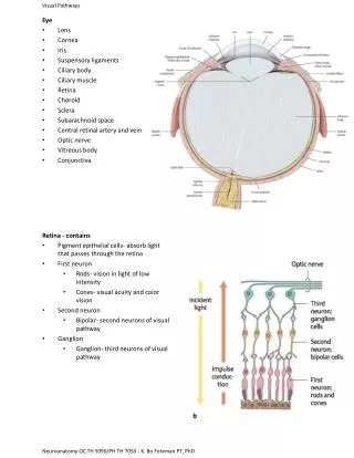
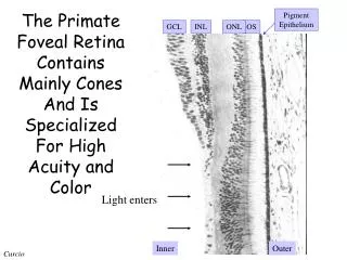
Light enters. The Primate Foveal Retina Contains Mainly Cones And Is Specialized For High Acuity and Color. Pigment Epithelium. GCL. INL. ONL. OS. Inner. Outer. Curcio. Retinal Cell Types Can Be Distinguished By Their Size And Branching Patterns (Dacey, 1993; Rodieck 1998).

E N D
Light enters The Primate Foveal Retina Contains Mainly Cones And Is Specialized For High Acuity and Color Pigment Epithelium GCL INL ONL OS Inner Outer Curcio
Retinal Cell Types Can Be Distinguished By Their Size And Branching Patterns(Dacey, 1993; Rodieck 1998)
Cell Size Within A Class Usually Increases With Eccentricity(Dacey, 1993; Rodieck, 1998) Parasol Dendritic field diameter (microns) Small bistratified Midget Retinal eccentricity (mm)
Multiple Retinal Pathways Exist, Having Very Specific Spatial Patterns of Cone Connections(Rodieck, 1998)
Dacey e-mail from April 22, 2003 • Inner and outer parasol cells • Inner and outer midgets • Small bistratified, • Large bistratified, • Inner and outer thornys • Dense thorny (this is an ON-OFF type that is broadly stratified across the ON-OFF border), • Large sparse monostratified – inner and outer • Giant monostratified (these are the photoreceptive ganglion cells - they stratify at the extreme inner and outer borders of the IPL but are NOT ON and OFF types and at the moment I have lumped them into a single physiological/anatomical grouping). There are also at least three other groups that we label from the colliculus that I have not seen consistently from the LGN (these probably include the ON-DS and the ON-OFF DS physiological groups).
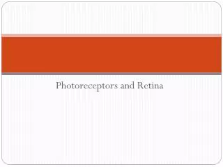
Cones Chemical synapse Electrical synapse Rod Circuitry(Kolb) Rods Hyperpolarizing rod photoreceptors depolarize rod bipolar cells Rod signals reach OFF-center ganglion cells
Rod bipolar AII amacrines The Retina Contains A Specialized Rod Pathway(Macaque, rod pathway; Massey in Rodieck Book)