BASIC VENTILATION
BASIC VENTILATION. Dr David Maritz. Mechanical ventilation. Emergency Med Clin N Am. 26 (2008) 849 - 862. Introduction. Emergency room-vs-ICU-vs-operating room Trouble shooting in ICU Terminology! Specific scenarios. Introduction. Why is the patient on the ventilator?

BASIC VENTILATION
E N D
Presentation Transcript
BASIC VENTILATION Dr David Maritz
Mechanical ventilation. Emergency Med Clin N Am. 26 (2008) 849 - 862
Introduction • Emergency room-vs-ICU-vs-operating room • Trouble shooting in ICU • Terminology! • Specific scenarios
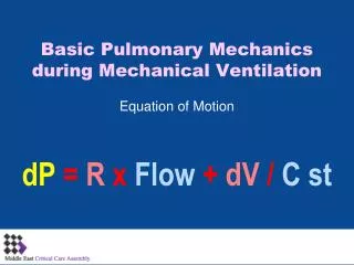
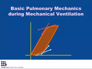
Introduction • Why is the patient on the ventilator? • Is the patient breathing spontaneously? • Who is doing the greater work of breathing? • Volume or pressure targeted strategy? • Dual controlled mode? • What is the set respiratory rate? • What is the total respiratory rate? • What is the set extrinsic / applied PEEP? • Is there intrinsic / auto PEEP? • What is the I:E ratio, flow rate, trigger mode? • What do the respiratory graphics indicate?
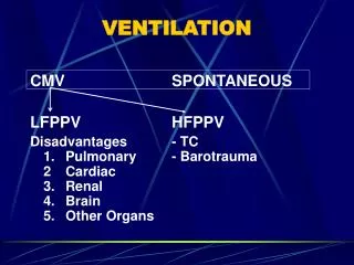
Introduction • Volume targeted ( volume cycled , volume assist / control) • Pressure targeted • Dual
Introduction • RR x Vt = MV • Intubated for airway protection • Septic / severe acidosis • ALI / ARDS • Other scanarios
Introduction • Adjust FiO2 • Extrinsic PEEP – offset loss of FRC • Caution in: • Elevated ICP • Unilateral lung process • Hypotension • Hypovolaemia • Pulmonary embolism
Introduction • Flow waveform – decelerate • Optimise recruitment • Trigger mode – detects pressure or flow gradient • Patient triggers ventilator • Too high – increased work • Too low – auto trigger • 1 – 3 cmH2O
Spontaneous breathing • Supported by pressure support ventilation (PSV) • Clinician sets FiO2 / PEEP • Patient sets RR / flow rate • VT dictated by PS / patient effort / lung compliance • Back up apnea rate
Volume targeted mode • Ventilator will generate necessary driving pressure to reach the targeted volume • Beware auto / intrinsic PEEP ( breath stacking) • Therefore progressive air trapping
Pressure targeted mode • Ventilator generates preset inspiratory pressure • Vt function of respiratory mechanics • Better pressure distribution • Any change in system compliance / resistance will affect Vt
Asthma / COPD • Volume depleted • Hyperinflation (auto-PEEP) • 8-10 breaths per minute • Decrease inspiratory time / increase expiratory time • Vt 6-7 ml/kg • Increase flow rate (80-100l/min) • Square wave form • Permissive hypercapnia • Sedation / paralyze • Sudden hypotension: • Disconnect fom ventilator • Tension pneumo
Acute lung injury / ARDS • ARDS: • PaO2/FiO2 < 200 • Bilat pulmonary infiltrates • Wedge presssure < 18mmHg • ALI: • PaO2 / FiO2 < 300 • Lung protection ventilation: • Vt 4-6ml/kg • Higher resp rates • Plateau pressures < 30cmH2O • Permissive hypercapnia • Volume targeted • Sedation / temp paralysis
Troubleshooting • Respiratory distress in ventilated patients: • Anxiety • Pain • Inadequate ventilator settings • ETT malfunction • Pulmonary parenchymal process • Extrapulmonary process • Tension pneumotghorax • Severe auto-PEEP • Stable – vs - unstable
Hemodynamically stable • Systematic approach • Focused history / exam • Check ventilator / circuit • Respiratory mechanics ( Peak and Plateau pressures) • CXR • Bedside ultrasound
Hemodynamically unstable • Remove from ventilator / hand ventilated 100% O2 (beware if high PEEP) • Severe auto-PEEP: • Do not hyperventilate • Disconnect from ventilator / compress chest • Tension pneumothorax: • Both sides! • Check settings / circuit / ETT etc • Reintubation – DIFFICULT AIRWAY
Noninvasive positive pressure ventilation in the emergency department. Emerg Med Clin N Am. 26 (2008) 835 - 847
Definition • CPAP a separate entity! • Continuous positive pressure • Tight fitting facemask • Spontaneous breathing • NPPV / NIPPV / Bilevel pressure • Inspiratory pressure (IPAP / inspiratory positive airway pressure) • End expiratory positive pressure (EPAP / expiratory positive airway pressure) • Breaths triggered by patient (back up rate)
Rationale • Avoid complications of invasive ventilation • Avoid ICU admissions • Reduce costs • Improve mortality
Advantages of NIV Preservation of airway defence mechanism Early ventilatory support Intermittent ventilation Patient can eat, drink and communicate Ease of application and removal Patient can cooperate with physiotherapy Improved patient comfort Reduced sedation requirements Avoidance of complications of intubation Ventilation outside hospital setting possible Disadvantages Mask is uncomfortable/claustrophobic Time consuming for medical and nursing staff Airway is not protected No direct access to bronchial tree for suction
Pathophysiology • CPAP – increases alveolar recruitement • = extrinsic PEEP and EPAP • Negates intrinsic PEEP ( auto PEEP / dynamic hyperinflation) • Increases intrathoracic pressure • NPPV / bilevel • IPAP = pressure support • Rest during inspiration
Indications • Acute exacerbations COPD • Asthma • Acute pulmonary oedema • Hypoxemic respiratory failure • Immunosuppressed patients • Do not intubate patients • Facilitation of weaning and extubation
Exacerbation COPD • Initiate early • Alongside with medical therapy
Asthma • Extrinsic PEEP offsets intrinsic PEEP
Acute cardiogenic pulmonary edema • CPAP and NPPV improve symptoms • Neither improves mortality • May decrease intubation rates
Hypoxic respiratory failure • Mixed data • Further studies needed
Feasibility • Very little data on safety • Failure of NPPV associated with: • GCS < 13 • RR > 20 after 1 hour • pH < 7.35 after hour
Initiation • No standard approach • High-low approach: • High IPAP (20-25cmH2O) • Low-high approach: • Low IPAP (8-10cmH2O) • EPAP 3-4cmH2O • Significant autopeep / PEEPi - EPAP 4-8cmH2O • Titrate FiO2 • Adjust EPAP
Summary • Reversible conditions • Bridging therapy • Close monitoring / follow up