LUNG CANCER
LUNG CANCER. ALOK SINHA Department of Medicine Manipal College of Medical Sciences Pokhara , Nepal. Epidemiology . One of commonest cause of cancer death Responsible for 25% of all cancer deaths More than 3 folds increase in cancer deaths since 1950 . Male: female - 2: 1


LUNG CANCER
E N D
Presentation Transcript
LUNG CANCER ALOK SINHA Department of Medicine Manipal College of Medical Sciences Pokhara, Nepal
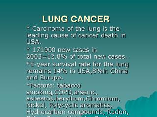
Epidemiology • One of commonest cause of cancer death • Responsible for 25% of all cancer deaths • More than 3 folds increase in cancer deaths since 1950
Male: female - 2: 1 • 8% cancer deaths in men and 4% of cancer deaths in women • Increasing in women • increasing smoking
Predisposing Factors 1. Cigarette smoking • Main predisposing factor • 90% smoking-related • Stopping smoking decreases the risk • Passive smoking can also put people at risk • High increases beyond 30 pack years 2.Exposure to Asbestos • Asbestos exposure + cigarette smoking particularly harmful
3.Other occupational exposures • Arsenic • preservation of wood • added to a semiconductor like transistors • in the tanning industry as a preservative • anti-friction additive in ball bearings • Chromium • manufacture of stainless steel • electroplating • preservation of wood • used as a yellow pigment • Nickel & other heavy metals • Used as alloys in many industries can also increase risk
4.Radiation exposure damages the DNA material within the cells and can also cause lung cancer 5.Radon(a radioactive gas) exposure can predispose to lung cancer. people living in areas with a high radon content are prone to increased incidences of a variety of cancers Levels high in basement
6. Pre existing non malignant lung diseases associated with increased lung cancer rates • Tuberculosis • Chronic obstructive pulmonary disease • Idiopathic pulmonary fibrosis
1. Non small cell lung cancer (NSCLC)75 -80% • Squamous cell carcinoma • Adenocarcinoma • Alveolar cell carcinoma • Large cell carcinoma 2. Small cell lung cancer (SCLC)20 - 25% 3. Miscellaneous • Carcinoid • Sarcoma • Unspecified lung cancer Oat Cell cancer
Non-small cell lung cancer (NSCLC) Squamous cell carcinoma • Commonest histological type • Centrally located • Slow growing • take several years to progress from a confined tumor into invasive cancer • Causes pneumonia atelectasis etc • Presents as a mass on CXR • May cavitate and look like a lung abscess • Response to chemotherapy is poor
(NSCLC contd) • Adenocarcinoma • Not always smoking-related • Are peripheral • Can occur in scar tissue • Can be a • lung primary • secondary from adenocarcinomas at other sites, especially if causing pleural infiltration • Have a worse prognosis than squamous cell cancer in all stages
Alveolar cell carcinoma • Rare • Can cause copious sputum production (bronchorrhoea) • Fluffy air space shadowing on CXR • Large cell carcinoma • an uncommon NSCLC • similar to that of adenocarcinoma
Small cell lung cancer (SCLC or Oat cell) • STRONGLY associated with smoking • More aggressive than NSCLC • Presents as central lesion with hilar & mediastinal invasion along with regional adenopathy • Rapidly growing -usually disseminated by the time of diagnosis(haematogenous spread) to • Liver • Bone & bone marrow • Brain • Adrenals, or elsewhere
Paraneoplastic syndromes like SIADH common (except PTH) • Surgery usually not appropriate • Chemo- and radiosensitive • Untreated – rapidly progressive and has a median survival of 6 weeks
Common Symptoms ? • Coughing – change in character in a smoker • Weight loss • Shortness of breath • Chest pain : often ill-defined and aching • Haemoptysis: sputum may be streaked with blood • Non-specific symptoms: fever, weakness, lethargy • Rarely • difficulty in swallowing • wheezing Monomorphic wheeze
Symptoms and signs Due to: • Local tumour effects • Metastatic tumour effects • Paraneoplastic manifestations
local tumour effects • Local involvement of the bronchus • Mediastinal spread • Compression of local nerves • Superior vena cava syndrome • Spread to the chest wall
Local involvement of the bronchus 1. Dry cough • Purulent if there is secondary infection • Change in the character of the 'regular' cough of a smoker 2. Haemoptysis (tumours arising in central bronchi) • Repeated episodes of scanty haemoptysis • blood-streaking of sputum in a smoker are highly suggestive of bronchial carcinoma and should always be investigated • Occasionally massive: large vessels invaded, -may be fatal
3. Bronchial obstruction Bronchial obstruction Complete obstruction Partial obstruction • Partial – hyperinflation • Complete – atelectasis • dyspnoea
4. Pneumonia • When the obstruction is insufficient to cause collapse • Recurrent pneumonia in the same sight • Delayed resolution of pneumonia 5. Lung abcess • Due to obstruction • Tumor undergoing necrosis 6. Wheeze/stridor due to airway obstruction • Monophonic or unilateral wheeze • failing to clear with coughing suggests presence of fixed bronchial obstruction
2. Mediastinal spread Oesophahgeal involvement causing dysphagia
3. Compression of local nerves • Left recurrent laryngeal nerve palsy • hoarse voice with 'bovine' cough • Phrenic nerve paralysis • unilateral diaphragmatic palsy • shoulder pain • dullness to percussion • paradoxical movement of abdominal wall • absent breath sounds at a lung base
4. Superior vena cava syndrome • Bilateral engorgement of the jugular veins • Distended veins over chest wall • Oedema of • Face • Neck • arms • conjunctivae
5. spread to the chest wall • Pleural pain • Pain due to intercostal nerve involvement • Pancoast tumor (superior sulcus tumor, or tumor of the pulmonary apex) • directly invade • sympathetic chain • Horner's syndrome (meiosis, ptosis, enopthalmos, anhidrosis) • brachial plexus • Cause weakness of small muscles of hand • C5/6, T1 motor loss • shoulder pain
Metastatic effects • Local metastasis • Blood born metastases
Local metastasis • Cervical/supraclavicularlymphadenopathy • common, present in 30% • easy site for diagnostic biopsy
Blood born metastases • C.N.S. involvement • focal neurological defects • epileptic seizures • personality change • Bone: pain & pathological fracture • Skin nodules • Liver: Jaundice • Lassitude, anorexia and weight loss usually indicate the presence of metastatic spread
Non-metastatic extrapulmonary manifestations(Para neoplastic syndrome) Endocrine • Inappropriate ADH secretion • causing hyponatraemia (SIADH) • Ectopic ACTH secretion – Cushings syndrome • Parathyroid hormone (PTH)-related peptides - hypercalcaemia • Carcinoid syndrome (Serotinin producing tumor causing diarrhoea brnchospasm) • Gynaecomastia
C.N.S. Symptoms precede those of primary tumour itself • Polyneuropathy • Myelopathy • Cerebellar degeneration • Myasthenia (Lambert-Eaton syndrome) • Limbic encephalitis • Limbic system: collection of structures at the base of the brain responsible for emotions and memory • Consists of Hippocampus, amygdala, anterior thalamic nuclei, limbic cortex • Symptoms: • memory loss, personality changes, anxiety or depression, neuropsychiatric disturbances, partial or generalized seizures, olfactory and gustatory hallucinations, sleep disturbances
Others Digital clubbing Hypertrophic pulmonary osteoarthropathy excessive proliferation of skin and bone at the distal parts of extremities, digital clubbing and periostosis of the tubular bones Nephrotic syndrome Polymyositis and dermatomyositis Eosinophilia
Differential diagnosis • Large no of conditions having • Cough & haemoptysis • Clubbing & Gynaecomazia • Pneumonic consolidation & collapse • Features due to secondaries & paraneoplastic syndrome – may be the presenting symptoms
1. CXR (PA and lateral) • location of lesion • pleural involvement, pleural effusion • rib destruction • intrathoracic metastases • mediastinallymphadenopathy • Normal CXR does not rule it out
COMMON RADIOLOGICAL PRESENTATIONS OF BRONCHIAL CARCINOMA 1.Unilateral hilar enlargement 2. Parenchymal shadows3. Lung, lobe or segmental collapse 4. Pleural effusion 5. Broadening of mediastinum, 6. enlarged cardiac shadow, 7. elevation of a hemidiaphragm 8. Rib destruction
2. Bronchoscopy • For centrally located tumors • Visualised directly using a flexible bronchoscope • Bronchio Alveolar Lavage can be done - cytology • Bronchial biopsies (and brush samples) can be taken for pathological examination • direct assessment can be made of operability judged by the proximity of central tumours to the main carina
3. Percutaneous needle biopsy (under CT or ultrasound guidance) • For peripheral lesions not accessible to the bronchoscope • In patients not fit enough for bronchoscopy • sputum cytology valuable diagnostic aid
. 4. Pleural biopsy • indicated in all patients with pleural effusions 5. Mediastinoscopy, thoracoscopy or thoracotomy • required to obtain a definitive histological diagnosis
In patients with metastatic disease diagnosis confirmed by • needle aspiration • biopsy of affected • lymph nodes • skin lesions • liver • bone marrow
Extent of primary tumour (T) • Regional lymph nodes (N) • Distant metastasis (M)
Possible only in 15% • In majority of cases (over 85%) surgery not possible due to • extensive spread • co-morbidity • Only palliative surgery possible for them • Best hope of long-term survival in slow growing tumors – squamous cell cancers limited to stage I or II
CONTRAINDICATIONS • Distant metastasis (M1) • Invasion of central mediastinal structures including heart, great vessels, trachea and oesophagus (T4) • Malignant pleural effusion • Contralateralmediastinal nodes (N3) • FEV1 < 0.8 litres (severe C.O.P.D.) • Severe or unstable cardiac or other medical condition
5-year survival rates of • Over 75% in stage I disease (N0, tumour confined within visceral pleura) • 55% in stage II disease (ipsilateralperibronchial or hilar node involvement)