Orthopedic Surgery
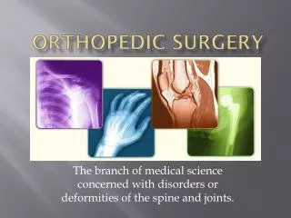
Orthopedic Surgery. The branch of medical science concerned with disorders or deformities of the spine and joints. Orthopedic Terminology “Position and Movement”. Abduction move a part away from body Adduction move a part toward the body Dorsiflexion bend or flex foot toward leg


Orthopedic Surgery
E N D
Presentation Transcript
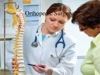
Orthopedic Surgery The branch of medical science concerned with disorders or deformities of the spine and joints.
Orthopedic Terminology“Position and Movement” • Abduction move a part away from body • Adduction move a part toward the body • Dorsiflexion bend or flex foot toward leg • Plantar flexion extend foot with toes pointed down (as when depressing the gas pedal) • Flexion to bend a part • Extension make a limb straight • Eversionturn outward • Inversion turn inward • Distal farthest away from point of origin • Proximal closest to point of origin • Medial nearest mid-line • Lateral away from the midline
Orthopedic Terminology“Position and Movement” • Valgus-abnormal displacement of part of a limb away from the midline of the body – distal from the affected joint – knees together • Varus- a deformity in which part of a limb is turned inward to an abnormal degree – distal from the affected joint – knees apart
Orthopedic Terminology • Acetabulum hollowed area of pelvis that receives head of femur • Acromioclavicular (AC) joint where clavicle joins acromion process of scapula • Arthritis inflammation of a joint • Arthrodesis surgical fusing or fixation of a joint • Arthroplasty surgical reconstruction of a joint • Arthroscopy visualization of a joint through an endoscope (diagnostic or operative) • Arthrotomy surgical incision into a joint • Articulation joint movement • Atrophy muscle wasting from lack of use • Bone marrow found in medullary canal of long bones and porosites of cancellous bone • Cartilage elastic, strong, dense connective tissue • Compact bone hard outer covering of bone • Cortical bone hard bone that forms shell of bones/acts as supporting structure • Condyle rounded part of a bone where ligaments articulate with adjacent bones • Curvature normal orabnormal bending
Orthopedic Terminology • Diaphysis shaft of a long bone • Dislocation displacement of a joint • Dysplasia abnormal tissue growth • Endosteum inside lining of bones where new bone forms (marrow) • Epiphysis two ends of a long bone • Exostosis bony growth arising from a bone’s surface • Fibroma tumor composed of fibrous or connective tissue • Foramen normal bone opening through which nerves, vessels, etc. pass • Foramen magnum occipital bone opening where spinal cord passes to vertebral column • Fossa shallow depression of a bone • Fracture break or crack of a bone • Hallux big toe • Implant implantation of graft (synthetic or tissue)
Orthopedic Terminology • Lamina flat layer or plate of a bone • Ligament connective tissue that joins bone surfaces • Malleolus rounded bone process (ankle) • Malunion faulty union of a fractured bone • Nonunion failure of fractured bone to unite • Osteogenesis origination/development of bone (ossification) • Osteomyelitis inflammation of bone tissue • Osteoporosis diminished calcium in a bone • Osteotomy surgical cutting of bone
Orthopedic Terminology • Pelvic Girdle bony structure that supports the trunk and provides attachment for the legs • Periosteum membrane surrounding bone (contains blood vessels) • Polydactylism more than normal number of digits • Scoliosis abnormal curvature of spine • Syndactylism webbing between digits • Synovial membrane lining of a joint capsule • Tendon fibrous tissue that connects muscle to bone • Traction force placed on bones or muscles to align or immobilize parts
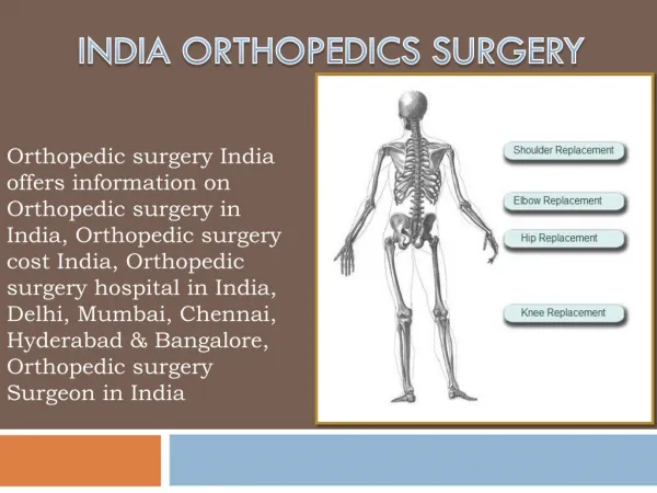
Primary purposes of orthopedic surgery • Repair, revision, reconstruction, reattachment or removal any of the 206 bones of the skeletal structure and surrounding tissue • Bones, joints and affected muscle tissue • Tendons, ligaments or cartilage • Accurately classify treatment • Axial skeletal procedures • Upper extremity procedures • Lower extremity procedures • Limb reattachment procedures • Amputation procedures
Purposes (continued) • Investigate, preserve and restore form and function to the musculoskeletal structures and associated tissues of the extremities and also the spine. Treatment depends on the type of injury and the duration of necessary immobility. • Stages of treatment • Investigation – diagnosis of structural issues • External • Internal • Preservation • Restoration • Types of treatment • Fracture management • Reduction • Immobilization • Rehabilitation • Corrective surgeries • Bone grafts • Implants • Internal and external fixation
Function of Skeletal System • Support • Protection • Movement • Storage • Hematopoiesis - the formation of blood cells in the living body (especially in the bone marrow)
Bone Histology • Bone is a type of connective tissue • 2 Types of Bone: • Dense/Compact Bone/ Corticalhard on outside/canal on insidecomposed of Haversian Units or Osteon • Spongy/Cancellous Bone
Bone Formation • Osteogenesis is bone formation • Two Types: • Intramembranous • Endochondral • More Terms: • Osteoblast :a cell from which bone develops • Osteocytes: a star-shaped cell, is the most abundant cell found in bone. They are osteoblasts that have completed their bone-forming function and have become trapped in new bone tissue, evolving into structural bone cells and is involved in the maintenance of that bone. A mature bone cell. • Osteoclasts: cells break down and assimilate bone. They are located in minute, bony chambers called lacuna.
Intramembranous Ossification • Sheets of primitive connective tissue form at site of future bone • Primitive connective cells collect around blood vessels in these layers • Connective tissue cells differentiate into osteoblasts, which deposit spongy bone • Osteoblasts become osteocytes when bony matrix surrounds them (lacunae) • Connective tissue on surface of each developing structure forms a periosteum • Osteoblasts on the inside of periosteum deposit compact bone
Endochondral Ossification • Masses of hyalin cartilage form models of future bones • Cartilage tissue breaks down/Periosteum develops • Blood vessels and differentiating osteoblasts from the periosteum invade the disintegrating tissue • Osteoblasts form spongy bone in space occupied by cartilage • Osteoblasts become osteocytes when bone matrix completely surrounds them • Osteoblasts beneath periosteum deposit compact bone around spongy bone
Relevant anatomy • Skeletal system – articulated skeleton comprised of 206 bones • Axial skeleton – skull, spine and ribs • Skull – includes cranial and maxillofacial bones • Cranial bones – 8 cranial bones • Facial bones – 13 facial bones • Middle ear bones – 6 middle ear bones • Mandible – one jaw bone • Hyoid bone • Vertebral column – 26 backbones • Verebral – 24 backbones: 7 cervical, 12 thoracic, and 5 lumbar • Sacrum – one sacrum bone • Coccyx – one tailbone • Thoracic cage – 25 thoracic bones • Sternum one cartilaginous bone that supports most ribs • Rib cage – 24 rib bones; 12 pair posteriorly attached to the spine
Relevant anatomy - Cervical • C-1 : a.k.a. Atlas: like Atlas man (Greek mythology), the bone supporting the skull. • C-2 Axis: bone that allows the head to pivot.
Appendicular Skeleton • The appendicular skeleton consists of 126 bones in the human body which make motion possible and protects the organs of digestion, excretion, and reproduction. • The word appendicular refers to an appendage or anything attached to a major part of the body, such as the upper and lower extremities. • The appendicular skeleton has four major regions: • Pectoral Girdles(4 bones) • Upper Limbs (60 bones) • Pelvic Girdle(2 bones) • Lower Limbs(60 bones)
Anatomy (continued) • Appendicular Skeleton • Upper extremities –shoulder, arm and hand bones of the appendicular skeletal system • Pectoral girdle – four pectoral or collar bones • Scapula – two posterior collar bones • Glenoid fossa • Coracoid process • Acromion process • Clavicle – two anterior collar bones • Upper limbs –arm, wrist and hand bones • Humerus • Radius • ulna
Anatomy (continued) • Lower extremities –hip and leg bones of the appendicular skeletal system • Pelvic girdle • Ilium • Pubis • Ischium • Lower limbs – leg and foot bones • Femur • Patella • Tibia • Fibula • Tarsals • Metatarsals • phalanges
Bone Marrow • Within the long bones are two types of bone marrow: red marrow and yellow marrow. • The yellow marrow is fatty tissue. • During starvation, the body uses the fat in yellow marrow for energy.
Bone Marrow • The red marrow of some bones is an important site for blood cell production. • Here all red blood cells, platelets, and white blood cells form in adults. • Red blood cells carry oxygen and nutrients to the body tissues. • Platelets help in blood clotting. • White blood cells help fight disease and infection.
Anatomy (continued) • Muscular anatomy • Neck muscles – sternocleidomastoid, platysma and trapezius • Torso muscles – deltoid, pectoralis, serratus anterior, latissimus dorsi, transverse abdominus, rectus abdominus and levator ani • Arm muscles – biceps brachii, triceps brachii, brachialis, brachioradialis, carpi, digitorium and pollicis • Leg muscles – gluteus, sartorius, quadriceps femoris, adductor, hamstring, gastrocnemius, tibialis anterior and digitorium, both flexor and extensor
Muscles • Functional unit of a muscle is the sarcomere • 3 Types: • Skeletalvoluntary/conscious movementstriated in appearancefound along-side skeletal system • Cardiacinvoluntary/unconscious movementfound only in myocardium of heart • Smoothinvoluntary/unconscious movementfound in the viscera
Knee anatomy Anterior Posterior
Foot Extensor digitorumlongus (EDL) – MUSCLE -The EDL extends or lift the toes
Bone composition types • Membranous bone – highly specialized connective osseous tissue that originally is membrane, then ossifies to bone • Cranial • Facial – maxilla (upper jaw), mandible (lower), nasal and lacrimal bones • Cartilaginous bone • Long bones • Flat bones • Irregular bones • Short bones • Sesamoid bones
Joint Classification • Functionallybased on degree of movement*synarthroses-no movement*amphiarthroses-slight movement*diarthroses-freely moveable • Structurallybased on type of connective tissue and type of joint cavity*fibrous-no movement, no joint cavity, dense fibrous connective tissue, synarthoses*cartiligenous-slight to no movement, can be synarthroses or amphiarthroses*synovial-joint cavity, diarthroses
Diarthroses Joints • The 6 types of diarthroses joints: • Ball-and-Socket • Condyloid • Saddle • Pivot • Hinge • Gliding
Ball-and-Socket Joint • The ball-shaped end of one bone fits into a cup shaped socket on the other bone allowing the widest range of motion including rotation. • Examples include the shoulder and hip.
Condyloid Joint • Oval shaped condyle fits into elliptical cavity of another allowing angular motion but not rotation.
Saddle Joint • This type of joint occurs when the touching surfaces of two bones have both concave and convex regions with the shapes of the two bones complementing one other and allowing a wide range of movement. • The only saddle joint in the body is in the thumb.
Pivot Joint • Rounded surfaces of one bone fit into a ring of one or tendon allowing rotation. • An example is the joint between the axis and atlas in the neck.
Hinge Joint • A hinge joint allows backward and forward movement in only one direction, much like a door opening and closing. • Examples • Knee joint • Elbow joint
Gliding Joint • Flat surfaces move against each other allowing sliding or twisting without any circular movement
Joints and surrounding tissue • Joints – points of articulation where movement between bones can occur • Axial skeleton • Skull • Cranial and facial sutures • Temporomandibular • Vertebral column • Atlanto-occipital • Intervertebral • Ribs and sternum • Sternoclavicular • sternocostal
Joints and surrounding tissue (continued) • Upper extremities • Pectoral girdle • Acromioclavicular • Shoulder (glenohumeral or humeroscapular) • Elbow • Hand • Wrist (radiocarpal) • Digit • Lower extremities • Pelvic girdle • Sacroiliac • Pubic symphysis • Hip • Knee (tibiofemoral and femoropatellar)
Joints and surrounding tissue (continued) • Tibiofibular (proximal and distal) • Ankle • Foot • Intertarsal • Metatarsophalangeal • Toe (interphalangeal) • Joint structure • Articular hyaline cartilage • Fibrous capsule • Fat pad • Articular joint disc • Ligaments – connecting bone to bone • Tendons – connect muscle to the bone • Synovial membrane and fluid
Joints and surrounding tissue (continued) • Joint articulation types • Synovial – allow free movement/have a joint cavity • Cartilaginous – allow little movement/no joint cavity • Fibrous – allow no movement/No joint cavity • Surrounding soft tissue • Circulatory – blood vessels • Peripheral nerves • Foramen • muscles
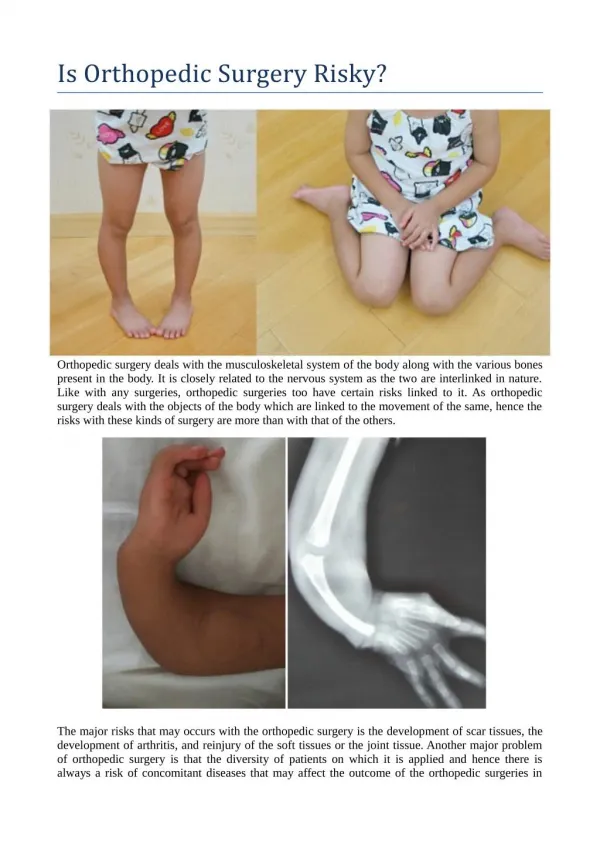
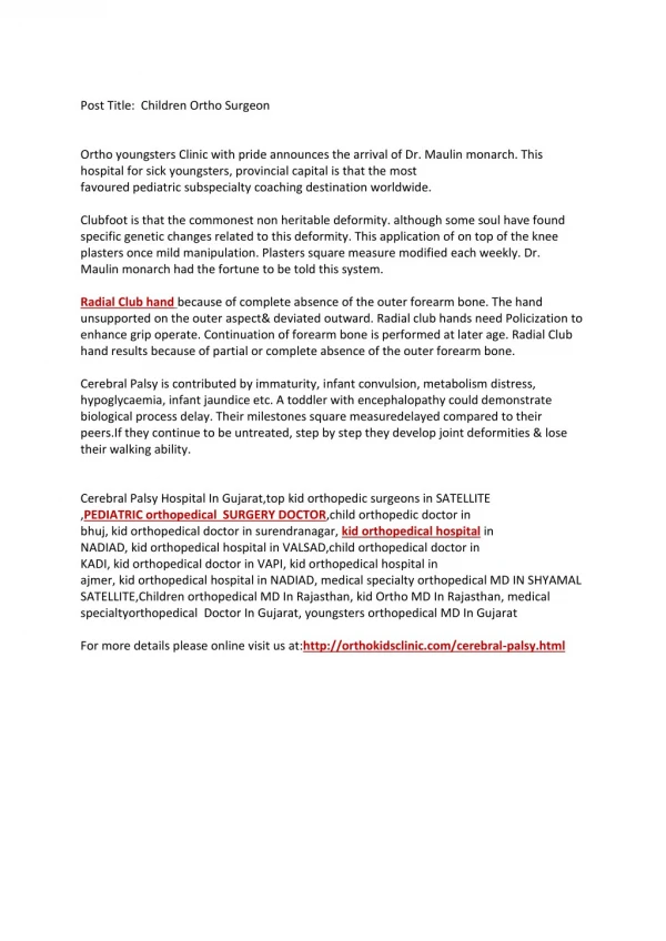
Pathology • Pathologic • Congenital • Dysplasia – abnormal tissue growth • Hip dislocation • Polydactylism • Scoliosis, kyphosis and lordosis – abnormal curvature of the vertebral column • Syndactylism – webbing between digits • Acquired disease • Arthritis – inflammation of a joint • Osteoarthritis (OA) • Rheumatoid arthritis (RA) • Bursitis – inflammation of the synovial fluid • herniation
Pathology (continued) • Infection • Osteomyelitis – inflammation of bone tissue • Calcium disorders • Rickets – vitamin D and calcium deficiency • Osteomalacia – soft bones • Osteoporosis – fragile and porous bones • Tumors • Osteochondroma – generally benign • Osteoma – benign tumor of the bone • Fibroma – composed of fibrous tissue • Osteosarcoma – malignant tumor of the bone • Myeloma – cancer in the bone marrow • Chondrosarcoma – tumors of the hyaline cartilage, often malignant • Volkmann’s contracture • Strain – stretching of joint tendons