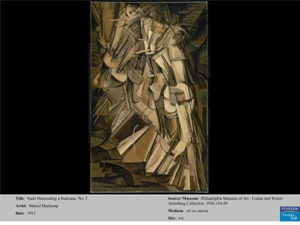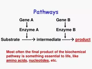
Comprehensive Overview of Motor Control Pathways in the Human Nervous System
Explore the intricate network of descending pathways and motor control centers in the spinal cord, brainstem, and cerebral cortex, detailing their organization and functions. Learn about the hierarchal and parallel arrangement of centers, the organization of motor neuron pools, and the different brainstem pathways influencing motor function. Gain insights into the role of aminergic pathways in modulating spinal neuron excitability. Discover the complexities of the pyramidal tract and cortical origins of motor control signals. Unravel the mysteries of muscle control and fine motor actions governed by the nervous system.

Comprehensive Overview of Motor Control Pathways in the Human Nervous System
E N D
Presentation Transcript
Motor control • Three levels of motor control: • Spinal cord • Brainstem • Cerebral cortex (motor <- premotor) • These centers are arranged in both hierarchical and parallel manner.
MN organization • Cell bodies organized into motor neuron pools – longitudal column spanning 1-4 segments • Two axis of organization • Proximal\distal rule: Proximal is medial • Flexor\extensor rule: Flexors are dorsal
IN organization • Segmental INs follow MN proximal/distal organization. • Propriospinal INs have axonal distribution that reflects their location: • Medial -> long • Distal -> short
2. Brainstem pathways (BSP) • Three descending systems: • Medial reaches ventromedial part of IZ proximal & axial musculature • Lateral dorsolateral part of IZ distal muscles. • Aminergic broad, diffuse innervation of SC.
MEDIAL PATHWAYS LATERAL PATHWAYS Tectum Red nucleus Medial Reticular formation Tectospinal Vestibular nuclei Reticulospinal Vestibulospinal Rubrospinal
Medial BSP • Vestibulospinal (M+L) - ipsilateral • Originates in the vestibular nuclei. • Balance and posture information • Reticulospinal (M+L) – ipsilateral • Originates in reticular formation nuclei • Excitation & inhibition of INs and MNs • Control of posture • Integrates cortical and vestibular information. • Cortico-reticulospinal pathways (M1 + PM). • Tectospinal– contralteral • Originates from the sup. Colliculus • Eye-head coordination. • Supply INs + long PNs in the ventromedial part of IZ
Lateral BSP • Rubrospinal tract – contralateral • Originates from the magnocellular part of RN • Reduced importance in higher primates
Medial vs. lateral BSPs • Medial BSP: • Phylogenetically oldest • Balance & posture control via proximal+distal muscles • Wide termination fields • Lateral BSP: • Wider motor spectrum • Reaching & object manipulation via distal musculature • Function overtaken by motor cortical areas.
Aminergic BSPs • Control the excitability of spinal neurons. • Ceruleospinal system: • NA • Locus ceruleos + pontinemedullary reticular formation. • Descend via ventral part of lateral column • Raphe-spinal system • Serotonin (5HT) • Raphe nuclei • Descend via ventral + lateral columns.
Axons reach the intermediate zone and motor nuclei Single neurons sends multiple collaterals which may span the entire cord. The raphespinal system modulate sensory transmission in the dorsal horn. Aminergic BSPs
Complex and precise motor actions depends on motor cortical signals. Motor cortical areas affect motor output via Direct corticospinal pathways (1*106) Indirect cortex-brainstem-spinal pathways (19*106) – CRetS, CRnS Most of the cortical command is further processed in subcortical-supraspinal centers. 3. Pyramidal tract
Morphology of PT • 60% of fibers from frontal lobe (motor areas) • 40% of fibers from parietal lobe (sensory areas) • 75% contralateral – dorsolateral column – lateral CST • 25% ipsilateral – ventral column – ventral CST
Ventral corticospinal Lateral corticospinal Red nucleus Medial brain stem pathways Pyramidal decussation Ventral corticospinal Lateral corticospinal
Cortical origin of PT • Pyramidal cells in layer V • In the M1 these are giant cells (Betz cells) • In PM these cells are smaller
Cortical organization of PT neurons • Intracellular recording of MNs and Ia-IN while stimulating M1 • Multiple patches project to single neurons • Single cortical site project to multiple neurons • Overlap between CS and CM projecting areas. • MUSCLE COLONIES
Ventral shift of CST • Corticospinal and corticobulbar pathwaty appear first in mammals • In higher mammals there is a dorsal-to-ventral shift of CST termination. • From sensory gating to motor control. • Direct CM contact facilitate dexterity.
Embryogenesis of CST: reduced overlap and synaptic clustering
Spinal termination of the PT • Large termination field • A single fiber probably innervates both INs and MNs