Vectors and EKG’s
810 likes | 1.21k Vues
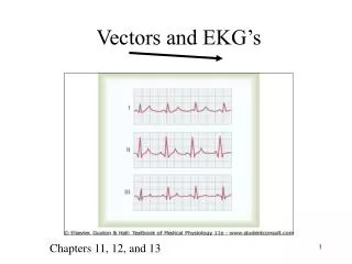
Vectors and EKG’s. Chapters 11, 12, and 13. Electrocardiogram (ECG). Depolarization wave passes through the heart and the electrical currents pass into surrounding tissues. Small part of the extracellular current reaches the surface of the body.

Vectors and EKG’s
E N D
Presentation Transcript
Vectors and EKG’s Chapters 11, 12, and 13
Electrocardiogram (ECG) • Depolarization wave passes through the heart and the electrical currents pass into surrounding tissues. • Small part of the extracellular current reaches the surface of the body. • The electric potential generated can be recorded from electrodes placed on the skin • An EKG is a comparison of two vectors • It compares the “heart vector” showing current flow on the heart with the reference, “recording lead vector” on the body. • (Non-invasive) • Heart Rate • Signal conduction • Heart tissue (enlarged) • Conditions (MI) • electrolyte and hormone imbalances
Vector diagrams • Vectors are used to describe depolarization and repolarization events • Vectors are arrows which show two things: • Direction or pathway (of charge spread) • Magnitude or size (amt of charge) • Vector analysis explains the waves on an EKG Q S
EKG is Extracellular Recording • Only looks at the charge on the outside of fibers! • Resting cell: outside positive • Depolarizing cell: outside negative • Repolarizing cell: outside positive • Depolarization: spread of surface neg charge • Repolarization: spread of surface positive charge • Vectors will always be positioned so that head of vector is in area of positive charge; tail is in area of negative charge. +++++++++++ ------------------ +++++++++++ ------------------ +++++++++++ ------------------ +++++++++++ ------------------
The following vectors represent the spread of negative charge during depolarization; Then the spread of positive charge during repolarization Rest No current flow, no vector.
- +
The atria would start to repolarize down and to the left, as the current continues downward to the ventricles We don’t detect this on the EKG, but what would the repolarizing vector look like? (review your specialized cells/contractile cells lecture!) +
Atria now have repolarized and now have positive surface charge again.
Meanwhile, as the atria are repolarizing...... We turn to the Depolarizing AV node These are small diameter fibers with few gap junctions; little or no detectable current flow
IV Septal Depolarization Moving down bundle of His; Current moves down R and L bundle branches from L toward R…why?
Through the thickness of the heart, from endo- , to myo-, to epicardium
Ventricles completely depolarized, negative surface charge No current No vector
Begin Ventricular Repolarization Spread of positive charge +
Rest End of cycle; No current flow, no vector.
Recording from Lead II Standard limb lead II
The Rules of Vectors Analysis • An EKG is a comparison of two vectors • It compares the “heart vector” with the reference “recording lead vector” on the body. • If the vectors run parallel (same direction) the pen moves upward from baseline • If the vectors run antiparallel (opposite direction) then the pen moves downward from baseline. • If the vectors are perpendicular, the pen remains on baseline. • If there is no current flow, the pen remains on baseline. • Each lead consists of two electrodes placed on the skin, with a voltmeter between them. • The voltmeter is attached to a pen, which travels over paper running at 25 mm/sec. This produces waves called an electrocardiogram.
- I + - - III II Einthoven’s Triangle Bipolar Limb Leads + +
Atrial depolarization Pen here V II T The heart vector is parallel to the lead, but how can you confirm?
Atrial depolarization - II • Draw a perpendicular line to the lead vector • Draw a line toward from the perpendicular vector toward your cardiac vector +
IV septal depol, from L to R II Anti-parallel! Pen deflects down Draw it!
Lateral walls depol II Draw it!
Depolarization complete; no current flow; pen returns to baseline II
Ventricular Repolarization complete; no current flow; pen on baseline II
Ventricular Repolarization complete; waiting to start all over again II End of one cardiac cycle
What does that tell you about the recording you obtain from each lead? • Each lead describes the events on the heart from “it’s own point of view” • Reading from several leads gives you different points of view about the same set of repeating events (depol, repol) • What if the recording lead was oriented this way? Use the words “down” or “up” to note the deflection compared to the five cardiac vectors above
Body Cross-section at Heart Level Heart V6 V5 V4 V3 V2 V1 12 Lead EKG’s • Read from each lead independently; one at a time over several heartbeats. • See what each lead shows. • 12 leads • 3 bipolar limb leads (I, II, III) • 3 augmented unipolar limb leads • (aVR, aVL, aVF) • 6 precordial leads (chest leads, V1-V6)
6 Leads- bipolar and augmented; all of these are in flat plane Augmented- Obtained by using the average voltage of any two points on skin as ground (neg pole) and reading from the third electrode (pos pole.)
Bipolar Leads and Einthoven’s Law • Lead I - The negative terminal of the electrocardiograph is connected to the right arm, and the positive terminal is connected to the left arm. • Lead II - The negative terminal of the electrocardiograph is connected to the right arm, and the positive terminal is connected to the left leg. • Lead III - The negative terminal of the electrocardiograph is connected to the left arm, and the positive terminal is connected to the left leg. • Einthoven’s Law states that the electrical potential of any limb equals the sum of the other two (+ and - signs of leads must be observed). Lead I LA – RA Lead III LL- LA Lead II LL- RA