Download
1 / 37
370 likes | 376 Vues
Prof. Hala Farawela Prof. of Clinical and Chemical Pathology Kasr El-Aini School of Medicine Cairo University. DR . Om ayman karrar -MD Clinical and Chemical Pathology.

E N D
Prof. Hala FarawelaProf. of Clinical and Chemical Pathology Kasr El-Aini School of Medicine Cairo University DR . Om ayman karrar -MDClinical and Chemical Pathology
Personal history: A Female patient, aged 70 yrs old ,Diabetic. Complaint: Abdominal pain, Fever , loss of weight, purities & weakness. Present history: The condition started 8 months ago by Abdominal pain , followed by fever , weakness& loss of weight. Abdominal examination: Palpable liver , no palpable LN . During the first 4months of the C/O she received symptomatic ttt without investigation.
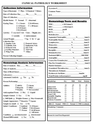
CBC Hb: 7.8 g/dlMCV:66 fl MCH:19.4 pg MCHC: 29 g/dl WBCs: 10 000/cmm S. Ferritin: 464 ng/ml Plts: 306 .000/cmm Differential count: Basophils: 0% Eosinophils: 30%Eosinophils: 30% (3000) Staff:3% Segmented:37% Lymphocytes:24% Monocytes: 6%
ESR: 130 mm/hr Sonar: Hepatomegaly Mild intrahepatic biliary dilatation. Hepatoduodenal LN suggesting Fasciolasis. Serology: for fasciola was negative
-ttt for Fasciola 2months No improvement Corticosteroides Negative PDGF α & β, Normal Karyotype, BCR-ABL1 was negative, Normal SPE, Mild increase IgE No Improvement Negative Hydroxyurea
After ttt with hydroxyurea , Eosinophilia ↓ markedly – Patient developed marked anemia , fever & persistent high ESR
touch smear cytology ++ plasma macrophages
BMB Diffusely fibrotic marrow infiltrated with Dorothy Reed Sternberg with background of macrophages , plasma cells, eosinophil & lymphocytes
Post contrast CT Chest, Abdomen & Pelvis done after revealed Mediastinal, Cervical , axillary, porta hepatis, panceratico duodenal ,para aortic & common iliac LN enlargement were seen.
Our case is Hodgkin's disease Patient run aggressive course and passed away.
Secondary Eosinophilia Parasitic Infectious Disease Allergic/ vasculitis Immunologic Drugs Non myeloid Malignancies Hodgkin's disease 2- Clonal Eosinophilia presence of histologic, cytogenetic, or molecular evidence of an underlying myeloid malignancy. CEL NOS. myeloid/lymphoid neoplasms with eosinophilia and mutations involving platelet-derived growth factor receptor α/β or fibroblast growth factor receptor 1 CML,MDS, CMML & SM. 3- Idiopathic eosinophilia Hypereosinophilic syndrome (HES) Mild 500-1500 cells/ µl Acquired Eosinophilia Moderate 1500-5000 cells/ µl Marked > 5000 cells/ µl