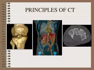
CT Scans - the principles
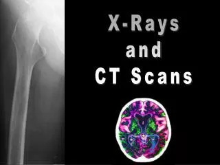
CT Scans - the principles. Craig Douglas. General Info. It is an X-ray imaging method Formation of an image is a distinct two stage process Principles described by Dr. Alan Cormack First system devised by Dr. Godfrey Hounsfield. The Process. Step 1- Scanning:


CT Scans - the principles
E N D
Presentation Transcript
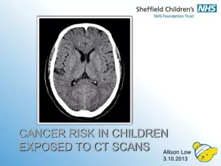
CT Scans - the principles Craig Douglas
General Info. • It is an X-ray imaging method • Formation of an image is a distinct two stage process • Principles described by Dr. Alan Cormack • First system devised by Dr. Godfrey Hounsfield
The Process • Step 1- Scanning: - A very thin X- ray beam is passed through the edges of a slice of body tissue - The beam is rotated around the body ( takes up to 15 seconds ) - Radiation penetrates to different degrees, depending on the density of the tissues they pass through ( dependent on the atomic numbers of the elements within the tissues ) - Electronic radiation detectors detect penetration • Step 2- Image Re-Construction
Still Scanners! • Third Generation -X-ray tube and radiation sensors both rotate in step with each other • Fourth Generation -X-ray tube rotates and radiation sensors form a continuous circle • No real indications of superiority
Image Formatting • A CT image appears to be a continuous display of a slice of tissue • But, No! • A CT scan is actually a matrix of individual elements
Image Formatting • The slice of tissue is formatted as an array of small volumes of elements, these are known as VOXELS • Each pixel on the monitor will represent a VOXEL • One pixel is displayed as uniform brightness, the elements within it are blurred together
Voxels and Pixels • The operator selects the number of pixels in an image, therefore this dictates the size of the voxels • As pixel number increases, voxel size decreases, allowing for better quality images.
CT Numbers • Each pixel can be represented by a CT number • The value is related to the physical density of the tissue in the corresponding voxel • Measured in Hounsfield Units • Water is the reference and assigned a value of zero
CT Numbers • Tissues more dense than water have a positive number • Same for opposites • CT Number =density (tis) – density (H2O) density (H2O) = ‘a’ X 1000
Image Display • Areas of high density, with high CT numbers appear white on the grey-scale • Areas of low density, with low CT numbers appear darker on the grey-scale
Windowing • Relationships between the CT numbers and the grey-scale can be adjusted by the system viewer • Viewer controls can set upper and lower CT number window limits • CT numbers > upper limit = white • CT numbers < lower limit = black • Easier distinction between soft tissues
Conversions 1000 0 -1000
Image Quality • Greater contrast sensitivity than plain radiography because: • All tissues are viewed directly, as opposed to looking at deep tissues obscured by superficial tissues • The relatively thin X-ray beam produces relatively less scatter • The ability to express a small range of CT numbers over the entire grey scale
Noise • This is a mottled appearance on CT • Its presence decreases the visibility of low contrast features • Produced due to the random manner in which x-ray photons are absorbed in the tissue • Decrease by: > x-ray dose (careful) or, > voxel size (poor)
Artifacts • Most commonly appear as streaks • Causes: - patient moving during scanning - metal objects in field of view
Radiation Dose • Measure absorbed radiation dose to the tissue within the scanned slice • Rad = 100 ergs per 1g tissue • Gray =1 joule per 1kg tissue • Represents radiation at a specific point, not total radiation applied • Different areas record different values
Dose Distribution • Direct exposure is limited to the tissue within the slice • A small amount will scatter to adjacent tissues • Total radiation energy increases with the no. of slices imaged and this must be considered • Generally even distribution, due to rotation of X-ray beam