Download
1 / 1
10 likes | 179 Vues
MICROPATTERNING OF BIOACTIVE GLASS NANOPARTICLES ON CHITOSAN MEMBRANES FOR SPATIAL CONTROLLED BIOMINERALIZATION. GISELA LUZ*, LUCIANO BOESEL Supervisors: Aránzazu del Campo and João F. Mano * gisela.luz@dep.uminho.pt. 9˚. Introduction

E N D
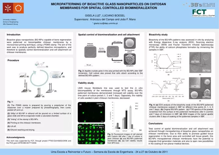
MICROPATTERNING OF BIOACTIVE GLASS NANOPARTICLES ON CHITOSAN MEMBRANES FOR SPATIAL CONTROLLED BIOMINERALIZATION GISELA LUZ*, LUCIANO BOESEL Supervisors: Aránzazu del Campo and João F. Mano * gisela.luz@dep.uminho.pt 9˚ Introduction Bioactive glass nanoparticles (BG-NPs) capable of bone regeneration were patterned on biocompatible chitosan membranes by a microcontact printing technique, using a PDMS stamp. The aim of this work was to produce perfectly defined bioactive micropatterns, and therefore to spatially control biomineralization and cell attachment on chitosan membranes. Spatial control of biomineralization and cell attachment Bioactivity study Bioactivity of the BG-NPs patterns was assessed in vitro by analyzing with Energy dispersive X-ray analysis (EDX), Scanning electron microscopy (SEM) and Fourier Transform Infrared Spectroscopy (FTIR) the ability of calcium phosphates formation by immersing the samples in SBF. SBF soaking Chitosanmembrane BG-NPspatterning Biomineralizedpattern Procedure CellCulture 50 μm Cellpattern (1) Fig. 2: Apatitecrystalsgrew in the area printed with the BG-NPs after SBF immersion. Cell culture also proved that cells attach according to the defined BG-NPs pattern. (6) (2) Viability study L929 mouse fibroblasts line was used to test the in vitro biocompatibility of the membranes through MTS assay. BG-NPs patterned membranes demonstrated increased cell viability over the time point of culture period (1, 3 and 7 days). Oppositely, the viability of cells seeded in plain chitosan membranes, decreased. (5) (3) (4) Fig. 1: (1) The PDMS stamp is prepared by pouring a prepolymer of the elastomer over a master prepared by photolithography, then cured, peeled off and cut . (2) 549μl of BG-NP in ethanol will be poured on a limited surface of a glass slide and left to evaporate inside a saturated chamber. (3) “Inking” of the stamp in BG-NPs. (4) Printing on the chitosan membrane. (5) Lift-off. (6) Ethanol washing and drying. Chitosan day 1 (a) Fig. 4: (a) EDX analysis of the bioactivity study of the BG-NPS patterned chitosan membranes soaked in SBF for different time points (0, 1, 3, 5 and 7 days). (b) Original BG-NPs pattern. (c) FTIR spectra of the powder scratched from the surface of the patterned membranes after 0 (control) and 7 days of immersion in SBF. (d) SEM images of the typical apatite clusters after 5 days of soaking of the patterned samples in SBF. Conclusions Total control of spatial biomineralization and cell attachment was achieved through micropatterning of bioactive glass nanoparticles on chitosan membranes. Due to their ability to promote guided tissue regeneration in the bone area and controlled soft tissue growth, this BG-NPs micropatterned chitosan membranes have the potential to integrate third generation materials and also to open new possibilities in the coating of non-planar medical devices. BG-NPs pattern day 1 (b) (c) Fig. 3: Fluorescent images of cell stained with Calcein AM after 1 day of culture in plain chitosan(a) and BG-NPs patterned membranes (b); (c) Cell viability results from MTS test. Acknowledgments This work was supported by FCT, through project PTDC/QUI/69263/2006 and the PhD grant SFRH/BD/45777/2008.


![DIGITAL DESIGN 3rd edition by MORRIS MANO Chapter 4 Combinational Logic Solutions to the Questions [1-35] DUYGU SA](https://cdn4.slideserve.com/966757/slide1-dt.jpg)