
Effects of Cylindrical Socket Design on Motion Loss in Transhumeral Prosthetics
This figure illustrates the cross-sections of a conventional transhumeral socket, demonstrating how the cylindrical design leads to a significant loss of motion during arm movement. The initial state shows the socket wall in relation to the humerus before force is applied. An upward force causes the humerus to shift, compressing the surrounding tissue, leading to a reduction in function. This motion loss occurs before the arm can effectively utilize the socket, indicating the importance of optimizing socket design for better rehabilitation outcomes.

Effects of Cylindrical Socket Design on Motion Loss in Transhumeral Prosthetics
E N D
Presentation Transcript
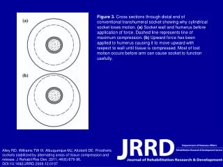
Figure 3. Cross sections through distal end of conventional transhumeral socket showing why cylindrical socket loses motion. (a) Socket wall and humerus before application of force. Dashed line represents line of maximum compression. (b) Upward force has been applied to humerus causing it to move upward with respect to wall until tissue is compressed. Most of lost motion occurs before arm can cause socket to function usefully. Alley RD, Williams TW III, Albuquerque MJ, Altobelli DE. Prosthetic sockets stabilized by alternating areas of tissue compression and release. J Rehabil Res Dev. 2011; 48(6):679-96.DOI:10.1682/JRRD.2009.12.0197