Download
1 / 41
410 likes | 954 Vues
ACUTE ABDOMINAL EMERGENCIES. Abdominal Anatomy and Physiology Abdominal pain and distress Abdominal conditions. Function of organs Digestion Stomach Small intestine Large intestine (colon) Liver Gallbladder Pancreas. Digestion Stomach: Hollow organ; expands as it fills with food

E N D
Abdominal Anatomy and Physiology • Abdominal pain and distress • Abdominal conditions
Function of organs Digestion • Stomach • Small intestine • Large intestine (colon) • Liver • Gallbladder • Pancreas
Digestion • Stomach: Hollow organ; expands as it fills with food • Small intestine: Hollow organ where food absorption takes place; Divided into 3 parts: Duodenum, jejunum, ileum • Large Intestine; hollow organ; removes water from waste products
Liver Bile secretion for breakdown of fats • Gallbladder Stores bile before release into the intestine • Pancreas Releases enzymes that breakdown food into absorbable molecules. Takes place in the small intestine
Reproductive • Endocrine Produces hormones ie insulin • Regulatory
Peritoneum • forms the lining of the abdominal cavity or the coelom — it covers most of the intra-abdominal (or coelomic) organs. It is composed of a layer of mesothelium supported by a thin layer of connective tissue. The peritoneum both supports the abdominal organs and serves as a conduit for their blood and lymph vessels and nerves.
The outer layer, called the parietal peritoneum, is attached to the abdominal wall. • The inner layer, the visceral peritoneum, is wrapped around the internal organs that are located inside the intraperitoneal cavity. • The potential space between these two layers is the peritoneal cavity; it is filled with a small amount (about 50 ml) of slippery serous fluid that allows the two layers to slide freely over each other.
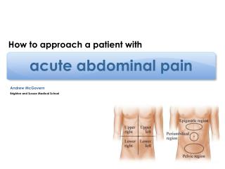
RUQ • Liver • Gall Bladder • Duodenum • Pancreas • Colon
Gall Stones • Hepatitis • Liver Disease • Pancreatitis • Appendicitis • Peforated Ulcer • AMI • Pneumonia
Left Upper Quadrant • Stomach • Spleen • Left lobe of Liver • Body of Pancreas • Left Kidney • Colon • Parts of Transverse and Descending Colon
Gastritis • Pancreatitis • AMI • Pneumonia
Gastritis: Inflamation of the lining of the stomach • Common causes Excessive alcohol consumption Prolonged use of NSAIDS such as Ibuprofen and ASA
Right Lower Quadrant • Cecum a pouch, connecting the ileum with the ascending colon of the large instestine. • Appendix • Right ovary and Fallopian tube • Right ureter
Appendicitis • Ruptured ectopic pregnancy • Pregnancy • Enteritis • PID • Ovarian cyst • Kidney stones • Abdominal abscess • Strangulated hernia
Enteritis Enteritis is an inflammation of the small intestine caused by a bacterial or viral infection. The inflammation frequently also involves the stomach (gastritis) and large intestine (colitis).
LLQ • Part of descending colon • Sigmoid colon • Left ovary and Fallopian tube
Ruptured ectopic pregnancy • Ovarian cyst • PID • Kidney stones • Diverticulitis • Enteritis • Abdominal abscess
Midline • Bladder infection • Aortic aneurysm • Uterine disease • Intestinal disease • Early appendicitis
Diffuse Pain The word "diffuse" means "widespread" and refers to pain that is more or less all over, or at least in many areas.
Pancreatitis • Peritonitis • Appendicitis • Gastroenteristis • Disecting/rupturing aortic aneurysm • Diabetes • Ischemic bowel • Sickle cell crisis
Visceral Pain • Dull and persistent Usually originating from solid organs • Intermittent, crampy, or colicky Pain comes from hollow organs
Parietal pain • Also called peritoneal pain • May be caused by internally bleeding • May be sharp and localized • May worsen when patient moves
Tearing pain • AAA tearing pain in the back Referred pain • Felt somewhere other than where it originates • MI-indigestion
Assessment and Care • Scene Size-up Protect yourself from vomit Odors Shock MOI
Initial Assessment LOC ABCs Signs of shock AMS Anxiety Pale Cool, moist skin Rapid pulse and respirations Position of patient O2
S A M P L E • O P Q R S T Time: How long have you had the pain Has it changed over time
Female patients • Where are you in your menstrual cycle? • Period late? • Vaginal bleeding? • If menstruating, is flow normal? • PMHx
Is pregnancy possible? Ectopic pregnancy is a priority pt., rapid transport.
Geriatric • Decreased ability to perceive pain • Medications for HTN or heart conditions that would prevent increased pulse when in shock
Beta Blockers Stimulation of β1 receptors by epinephrine induces a positive chronotropic(changes heart rate) and intropic(force of muscular contractions) effect on the heart and increases cardiac conduction velocity and automaticity. Beta Blockers Atenolol Metoprolol
Physical Exam of the Abdomen • Inspect Distension Bloating Discoloration Protrusions
Palpate • Localize pain prior to palpating palpate that area last • Observe for guarding • Carefully palpate a mass ONCE VS Serial vs
Care • ABCs • O2 • Transport decision • Position of comfort • Ongoing assessment q 5 min. • Alert for vomiting; suction • Calm • Nothing by mouth • AMS or unresponsive; left lateral recumbent • Elevate legs for shock
Appendicitis • Nausea and sometimes vomiting • Persistent pain RLQ Gallstones • Sudden epigastric/RUQ pain • May rotate to shoulder or back • May worsen by eating food high in fat
Pancreatitis • Pain may radiate to back and shoulders • Can be present with signs of shock Internal bleeding • Digestive tract; coffee ground emesis • Rectal; black, tarry stools • Paritoneal cavity; abd pain and tenderness
AAA • Sharp, tearing pain radiating to the back • Shock • Difference between femoral and pedal pulses Hernia • Painful protrusion Kidney stones • Severe flank pain radiating to anterior groin • Nausea and vomiting