Download
1 / 23
230 likes | 254 Vues
Learn about DNA arrays, their fabrication, and their application in disease detection. Explore the steps involved in microarray fabrication and analyze gene expression patterns in various diseases. Presented by Vigneshwaran Mani.

E N D
DNA arrays for early disease detection Chem 395 Bioanalytical chemistry Instructor Prof.James F.Rusling Presented by Vigneshwaran Mani
Outline • What is a Microarray? • Types of Microarray • Steps involved in Microarray fabrication • What happens to the Genes in disease state? • Application of Microarray • Summary
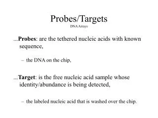
Why Microarrays????? What is a Microarray? A microarray is a spatially ordered, minituarized arrangement of multitude of immobilized reagents • Small volume- miniaturization • High throughput analysis • Large information generated • Less time required to analyze
Types of Microarrays • cDNA and Oligonucleotide Microarrays Probe: ssDNA,Oligonucleotide Target: ssDNA,ssRNA Principle: Hybridization • Protein Microarrays Probe: Antibody Target: Antigen Principle: peptide chemistry • Others Tissue arrays
Gene expression Nucleus RNA POLYMERASE RIBOSOMES Cytoplasm
cDNA ARRAYS Marketed By: Agilent technologies
Steps Involved in Microarrays • Microarray printing • Hybridization • Detection
100-10000 spots Glass slide used as substrate DNA is attached covalently to glass slide 96-384 well microtitre plates used Spot Volume 0.25-1nl. Spot size 100-150µm in diameter 1,2,3-X,Y,Z direction 4-print head 5-glass slide 6-Microtitre plate 7-Distilled water 8- Drying DNA-Microarray printing
Hybridization mRNA Control mRNA Diseased C U G C U G A A Reverse transcriptase Reverse transcriptase C C T G T G A A cDNA Fluorescent labeling Fluorescent labeling Cy3 Cy5 C C T G T G A A Hybridize target to microarray
Detection: The slide is scanned twice -Once to measure red intensity -Once to measure green intensity The images are overlayed to produce one image
Scanning M = logR/G = logR – logG • M<0: gene is over-expressed in green-labeled sample compared to red-labeled sample. • M=0: gene is equally expressed in both samples. • M>0: gene is over-expressed in red-labeled sample compared to green-labeled sample.
What happens to Genes in (Cancer) Disease State • Certain genes undergo overexpression. • No. of copies of particular genes may increase. • Gene mutation.
Gene is overexpressed in a certain disease state, More cDNA(target) will hybridizes to probe, as compared to control cDNA, In turn, the spot will fluorescence red with greater intensity than it will be with green. Expression patterns of various genes is characterized involved in many diseases, Compare expression pattern of the gene from the individual with the expression pattern of a known disease. Changes in gene expression levels
Genomic gains and losses • Number of copies of a particular target gene has increased. • Large amount of (Disease) sample DNA will hybridize to those spots on the microarray compared to (normal) control DNA hybridizing to those same spots. • Those spots containing the sample DNA will fluoresce red with greater intensity than they will fluoresce green, indicating that the number of copies of the gene involved in the disease has gone up.
Gene Expression profile analysis in Human hepatocellular carcinoma by cDNA microarray • Eun Jung Chung,Young Kwan Sung,MohammadFarooq,Younghee Kim, Sanguk Im,Won Young Tak,Yoon Jin Hwang, Yang Il Kim, Hyung Soo Han, Jung-Chul Kim, and Moon Kyu Kim.,Mol.Cells,Vol.14,pp382-387,2002
HCC (Hepatocellular carcinoma) • Primary liver cancer (HCC) • Somatic mutations and activation of certain oncogenes. • These events Lead to expression changes in genes. • cDNA arrays are used to analyze expression patterns
cDNA arrays Computer analysis 3063 human cDNA 8 different samples of HCC
Up-regulated genes in HCC • Galectin-3 • Serine/threonine kinase • Fibroblast growth factor receptor • Ribosomal protein L35A Down-regulated genes in HCC • mRNAs of Nip3 • Decorin • Insulin-like growth factor binding protein-3 Gene expression patterns of 8 hepatocellular carcinomas.The genes were primarily classified into three groups, based on their clustering pattern.
Summary • Data can be generated in a high throughput, parallel fashion. • Less time required for analysis. • If gene expression data is already known for a certain disease. Then we can compare the gene expression data of the individual with the known, and predict the disease
References • David J. Duggan, Michael Bittner, Yidong Chen, Paul Meltzer & Jeffrey M. Trent, Nature genetics supplement, volume 21, january 1999. • Sunil R. Lakhani, Michael J.O’hare & Alan ashworth, Nature medicine,volume 7,number 4,april 2001. • Vivian G. Cheung, Michael Morley, Francisco Aguilar, Aldo Massimi,Raju Kucherlapati & Geoffrey Childs, Nature genetics supplement,volume 21,january 1999. • http://www.ncbi.nlm.nih.gov/
Acknowledgement • Prof. James F. Rusling • Chem 395 students