Download
1 / 45
450 likes | 613 Vues
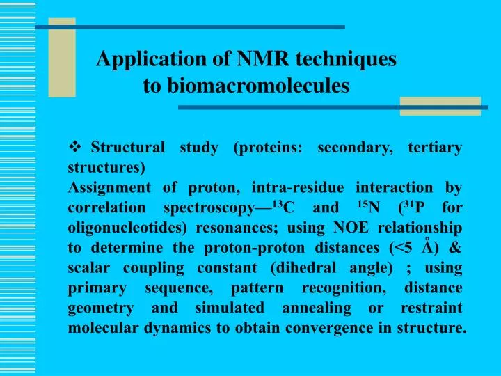
Application of NMR techniques to biomacromolecules. Structural study (proteins: secondary, tertiary structures)

E N D
Application of NMR techniques to biomacromolecules • Structural study (proteins: secondary, tertiary structures) • Assignment of proton, intra-residue interaction by correlation spectroscopy—13C and 15N (31P for oligonucleotides) resonances; using NOE relationship to determine the proton-proton distances (<5 Å) & scalar coupling constant (dihedral angle) ; using primary sequence, pattern recognition, distance geometry and simulated annealing or restraint molecular dynamics to obtain convergence in structure.
Application of NMR techniques to biomacromolecules • Dynamics study: from relaxation rate enhancement to deduce exchange rate and diffusion coefficient; protein folding using proton/deuterium exchange measurements and stopped-flow, and quenching, photo-activation • Chemical exchange and binding study—transferred NOE; relaxation enhancement by paramagnetic species (e.g. Mn2+) or spin labels
References: 1.A. Abragam, Principles of Nuclear Magnetism, Clarendon Press, Oxford, 1961. 2.K. Wüthrich, NMR of Proteins and Nucleic acids, John Wiley & Sons, N.Y., 1986. 3.I. Solomon, Phys. Rev. 99, 559-565, 1955. 4.J. Cavanagh, W.J. Fairbrother, A.G. Palmer III and N.J. Skelton, Protein NMR Spectroscopy: Principles and Practice, Academic Press, San Diego, 1996. 5.J.N.S. Evans, Biomoleculae NMR Spectroscopy, Oxford University Press, Oxford, 1995.
Dynamics: Spectral density function J(w)=2/5[τc/(1+ w2τc2)] whereτc is the correlation time which is a function of temperature, solvent viscosity and molecular size.
T1-1 =(3/2)g4ћ2I(I+1)[J(1)(wI)+J(2)( 2wI)] T2-1 =g4ћ2I(I+1)[3/8J(0)(0)+15/4J(1)( wI)+3/8 J(2)( 2wI)] Longitudinal and transverse relaxation by Scalar or J coupling between interacting nuclei (spin number I) Spin-spin coupling between a pair of nuclei via electrons in the bonds between them is the mechanism for J coupling. This results in a splitting of the resonance. It is an important basis of correlation spectroscopy.
The strategy for solving three-dimensional structure of biological macromolecules on the basis of NMR data
References: C.R. Cantor and P.R. Schimmel, Biophysical Chemistry, W.H. Freeman and Co., New York, 1980. R.F. Steiner and L. Garone, The Physical Chemistry of Biopolymer Solutions, World Scientific Co. , Singapore, 1991.
Surface Plasma Resonance: refractive index (RI) as a function of medium density; analyte immobilized on e.g. dextranwhich is bound to a gold surface; binding of substrate to analyte results in RI change; can be used in kinetics and binding studies
Sensor chip technology The sensor chip consists of a glass surface, coated with a thin layer of gold. This forms the basis for a range of specialized surfaces designed to optimize the binding of a variety of molecules. Dextran Analyte Gold Glass
T20 interation with gp41 N-terminal peptide gp41(516-566) Ka= 5.66e6 ; Kd=1.77e-7
Circular dichroism—to measure the secondary structures of proteins Linearly polarized light can be decomposed into right- and left-hand (RH and LH) circularly polarized light; when the polarized light is absorbed by optically active compounds, there may be a difference in the absorbance between RH and LH polarized lights, and thus circular dichroism. Proteins with regular secondary structures exhibit distinct pattern of CD spectra which ca be used to deduce the secondary structures of the proteins.
Effect of an optically active absorbing sample on incident linearly polarized light. All drawings show the electric field vector viewed along the direction of light propagation. Points 1 through 5 correspond to equal (increasing) time intervals. (a) Incident linearly polarized light. (b) Elliptically polarized light produced by passing the incident light through an optically active sample. (c) Resolution of linearly polarized light into individual right-hand and left-hand circularly polarized components. (d) Effect of an optically active sample on the two circularly polarized components. The sum of measurements made with these two separate components must be identical to the result obtained in part b.
References: J.R. Lakowicz, Principles of Fluorescence Spectroscopy. Plenum Press,New York, 1999.
Fluorescencespectroscopy: • Membrane binding study—emission wavelength and intensity change (NBD); fluorescence quenching—Trp and acrylamide • Self-assembly of biomacromolecules—self-quenching of fluorophore, e.g. rhodamine • Membrane fusion—fluorescence resonance energy transfer (FRET) between, e.g. NBD and Rhodamine-labeled phospholipids
Pathways for production and deexcitation of an excited state
D A Spectral overlap for fluorescence resonance energy transfer (RET)
The excitation wavelength was set at 467 nm; Symbols: • — , 0.04 mM NBD-WT alone; • ….. , a mixture of 0.04 mM NBD-WT and 0.04 mM Rh-WT; • - - , a mixture of 0.04 mM NBD-WT and 0.08 mM Rh-WT; • -.-. , a mixture of 0.04 mM NBD-WT and 0.12 mM Rh-WT Fluorescence energy transfers dependence on gp41 fusion peptide acceptor concentration