Download
1 / 42
470 likes | 820 Vues
Endocrine Glands & Hormones. BY ABDUL SAMIK DEPARTMENT OF VETERINARY REPRODUCTION, FACULTY OF VETERINARY MEDICINE, UNAIR. Learning Objectives: To understand what the reproductive endocrine glands and hormones are. To understand the characteristics and functions of these hormones.

E N D
Endocrine Glands & Hormones BY ABDUL SAMIK DEPARTMENT OF VETERINARY REPRODUCTION, FACULTY OF VETERINARY MEDICINE, UNAIR • Learning Objectives: • To understand what the reproductive endocrine glands and hormones are. • To understand the characteristics and functions of these hormones. • To understand how the concentrations of these hormones in the blood are controlled. • To understand the mechanism of action of the reproductive hormone
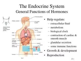
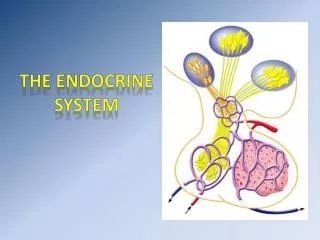
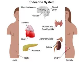
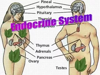
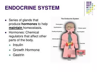
DEFINITION OF HORMONE A chemical substance secreted by an endocrine gland or group of endocrine cells that acts to control or regulate specific physiological processes, including growth, metabolism, and reproduction. Most hormones are secreted by endocrine cells in one part of the body and then transported by the blood to their target site of action in another part, though some hormones act only in the region in which they are secreted.
Hormone are classified according to : A. Mechanism of hormone action : • Reproduction hormone • Metabolic hormone • B. Biochemical structure: • 1. Peptide - Few - Several amino acids • Protein - Long chains of amino acids • Glycoprotein - Protein hormone with carbohydrate molecules • Steroid : derived from cholesterol • Fatty acid: derived from arachidonic acid • Amines : derived from tyrosine or tryptophan
HORMONE ACTION PROTEIN HORMONE STEROID HORMONE
Gonadotropin-Releasing Hormone • GNRH stimulates the synthesis and secretion of the gonadotropins, follicle-stimulating hormone (FSH) and luteinizing hormone (LH). • These processes are controlled by the size and frequency of GNRH pulses, as well as by feedback from androgens and estrogens. • GNRH secretion is pulsatile in all vertebrates, and is necessary for correct reproductive function. Thus, a single hormone, GNRH, controls a complex process of follicular growth, ovulation, and corpus luteum maintenance in the female, and spermatogenesis in the male • GnRH is considered a neurohormone, a hormone produced in a specific neural cell and released at its neural terminal. • A key area for production of GNRH is the preoptic area of the hypothalamus, that contains most of the GNRH-secreting neurons. • GnRH is secreted in the hypophysial portal bloodstream at the median eminence. • The portal blood carries the GnRH to the pituitary gland, which contains the gonadotrope cells
Gonadotropin-releasing Hormone (GnRH) • Gonadotropin-releasing hormone (GnRH) plays a key role in the regulation of the reproductive system. • GnRH has a similar structure in all animals. It is a decapeptide, meaning that it consists of a chain of 10 amino acids. • GnRH acts primarily to stimulate the anterior pituitary gland to synthesise and secrete the gonadotropins FSH and LH • It exerts three principal actions on the anterior pituitary gland: • Synthesis and storage of gonadotropins • Activation: the movement of gonadotropins from the reserve pool to a pool ready for direct action • Immediate release (direct secretion) of gonadotropins
The Posterior Lobe of The Pituitary • The posterior lobe of the pituitary releases two hormones, both synthesized in the hypothalamus, into the circulation. • VasopressinVasopressin is a peptide of 9 amino acids. • It is also known as arginine vasopressin (AVP) and the antidiuretic hormone (ADH) • Vasopressin acts on the collecting ducts of the kidney to facilitate the reabsorption of water into the blood. • This it acts to reduce the volume of urine formed (giving it its name of antidiuretic hormone). • OxytocinOxytocin is a peptide of 9 amino acidsIt acts on certain smooth muscles: • stimulating contractions of the uterus at the time of birth; • stimulating release of milk when the baby begins to suckle. • Oxytocin is often given to prospective mothers to hasten birth. Oxytocin also acts on the nucleus accumbens and amygdala in the brain where it enhances: • bonding between males and females after they have mated; • bonding between a mother and her newborn;
Control of Oxytocin Secretion • The most important stimulus for release of hypothalamic oxytocin is initiated by physical stimulation of the nipples or teats. • The act of nursing or suckling is relayed within a few milliseconds to the brain via a spinal reflex arc. • These signals impinge on oxytocin-secreting neurons, leading to release of oxytocin. • A number of factors can inhibit oxytocin release, among them acute stress. • For example, oxytocin neurons are repressed by catecholamines, which are released from the adrenal gland in response to many types of stress, including fright.
Another well-studied effect of steroid hormones is the marked increase in synthesis of uterine (myometrial) oxytocin receptors late in gestation, resulting from increasing concentrations of circulating estrogen • Both the production of oxytocin and response to oxytocin are modulated by circulating levels of sex steroids. • The burst of oxytocin released at birth seems to be triggered in part by cervical and vaginal stimulation by the fetus, but also because of abruptly declining concentrations of progesterone.
The Anterior Lobe of the Pituatary Thyroid Stimulating Hormone (TSH) • TSH (also known as thyrotropin) is a glycoprotein consisting of: a beta chain of 112 amino acids and • an alpha chain of 89 amino acids. The secretion of TSH is • stimulated by the arrival of thyrotropin releasing hormone (TRH) from the hypothalamus. • inhibited by the arrival of somatostatin from the hypothalamus. • TSH stimulates the thyroid gland to secrete its hormone thyroxine (T4). • A deficiency of TSH, or mutant TSH receptors, have also been implicated as a cause of osteoporosis.
Follicle-Stimulating Hormone (FSH), MW 32 kDa • FSH is a heterodimericglycoprotein consisting of the same alpha chain found in TSH (and LH) • a beta chain of 115 amino acids, which gives it its unique properties. • Synthesis and release of FSH is triggered by the arrival from the hypothalamus of gonadotropin-releasing hormone (GnRH). The effect of FSH depends on one's sex FSH in females • In sexually-mature females, FSH (assisted by LH) acts on the follicle to stimulate it to release estrogens. FSH in males • In sexually-mature males, FSH acts on spermatogonia stimulating (with the aid of testosterone) the production of sperm.
Luteinizing Hormone (LH), MW 30 kDa • LH is synthesized within the same pituitary cells as FSH and under the same stimulus (GnRH). • It is also a heterodimericglycoprotein consisting of the same 89-amino acid alpha subunit found in FSH and TSH (as well as in chorionic gonadotropin); • a beta chain of 115 amino acids that is responsible for its properties. • The effects of LH also depend on sex. LH in females • In sexually-mature females, a surge of LH triggers the completion of meiosis I of the egg and its release (ovulation) • stimulates the now-empty follicle to develop into the corpus luteum, which secretes progesterone LH in males • LH acts on the interstitial cells (also known as Leydig cells) of the testes stimulating them to synthesize and secrete the male sex hormone, testosterone. • LH in males is also known as interstitial cell stimulating hormone (ICSH).
Prolactin (PRL), MW 24 Da • Prolactin is a protein of 198 amino acids. During pregnancy it helps in the preparation of the breasts for future milk production. After birth, prolactin promotes the synthesis of milk. Prolactin secretion is • stimulated by TRH • repressed by estrogens and dopamine. ACTH — the adrenocorticotropic hormone • ACTH is a peptide of 39 amino acids. • ACTH acts on the cells of the adrenal cortex, stimulating them to produce : • glucocorticoids, like cortisol • mineralocorticoids, like aldosterone • androgens (male sex hormones, like testosterone • in the fetus, ACTH stimulates the adrenal cortex to synthesize a precursor of estrogen called dehydroepiandrosterone sulfate (DHEA-S) which helps prepare the mother for giving birth. • Production of ACTH depends on the intermittent arrival of corticotropin-releasing hormone (CRH) from the hypothalamus.
Growth Hormone (GH) • Growth hormone (GH; also called somatotropin) is a protein of 191 amino acids. • The GH-secreting cells are stimulated to synthesize and release GH by the intermittent arrival of growth hormone releasing hormone (GHRH) from the hypothalamus. • GH promotes body growth by: • binding to receptors on the surface of liver cells • this stimulates them to release insulin-like growth factor-1 (IGF-1; also known as somatomedin) • IGF-1 acts directly on the ends of the long bones promoting their growth • Alpha Melanocyte-Stimulating Hormone (α-MSH)
The ovaries release female sex hormonesincluding: • estrogen • Progesteron • In order to begin the ovulation process, the ovaries begin producing less estrogen. • This drop in estrogen signals your brain to release a special hormone, called gonadatropin-releasing hormone (GnRH). • Inhibin is a peptide that is an inhibitor of FSHsynthesis and secretion,[1] and participates in the regulation of the estrus cycle • Inhibin contains an alpha and beta subunit linked by disulfide bonds • In both females and males, inhibin inhibits FSH production and GnRH release in the anterior pituitary • FSH stimulates the secretion of inhibin from the granulosa cells of the ovarian follicles in the ovaries. In turn, inhibin suppresses FSH • Inhibin secretion is diminished by GnRH, and enhanced by insulin-like growth factor-1 (IGF-1).
In males • In men, it is a hormone that inhibits FSH by negative feedback. • It is secreted from the Sertolicells,located in the seminiferous tubules inside the testes. • Androgens stimulate inhibin production • this peptide may also help to locally regulate spermatogenesis Activinis a related peptide that counteracts inhibin • The release of GnRH, in turn, triggers the production of follicle-stimulating hormone (FSH), which actually gets your eggs maturing. • So, without your ovaries' hormone production, you wouldn't have any eggs to release. • The ovaries also help to develop your secondary sex characteristics
Placental Hormones Sex steroids Progesteron • Progestins, including progesterone, have two major roles during pregnancy: • Support of the endometrium to provide an environment conducive to fetal survival • Suppression of contractility in uterine smooth muscle. This is often called the "progesterone block" on the myometrium. Toward the end of gestation, this myometrial-quieting effect is antagonized by rising levels of estrogens, thereby facilitating parturition. Estrogens • Two of the principle effects of placental estrogens are: • Stimulate growth of the myometrium and antagonize the myometrial-suppressing activity of progesterone. In many species, the high levels of estrogen in late gestation induces myometrial oxytocin receptors, thereby preparing the uterus for parturition • Stimulate mammary gland development. Estrogens are one in a battery of hormones necessary for both ductal and alveolar growth in the mammary gland.
Natural estrogen : • Estradiol (C18H26O2) • Estron (C18H25O2) • Estriol (C18H27O3) Estrogen from plant (fitoestrogen) : • Coumestrol • Genisten • Zeranol Non steroid estrogen : • Diethylstilbestrol (DES)
human Chorionic Gonadotropin(hCG)MW 40 kDa • This hormone is produced by fetal trophoblast cells. • It binds to the luteinizing hormone receptor on cells of the corpus luteum, which prevents luteal regression. • Thus, hCG serves as the signal for maternal recognition of pregnancy Placental lactogen, MW 22-23 kDa • The functions of placental lactogens are thought to modulate fetal and maternal metabolism, perhaps mobilizing energy substrates for fetal use. • In some species they have been shown to stimulate function of the corpus luteum, and to participate in development of the mammary gland prior to parturition.
Relaxin, MW 5.700 Da • Relaxin is a hormone thought to act synergistically with progesterone to maintain pregancy. • It also causes relaxation of pelvic ligaments at the end of gestation and may therefore aid in parturation. • In some of the species in which relaxin is known to be produced, it is produced by the placenta, while in others, the major source is the corpus luteum. • In some species, relaxin is produced by both the corpus luteum and placenta • Structurally, relaxin is a heterodimer of two peptide chains of 24 and 29 amino acids that are linked by disulfide bridges and it appears related to insulin
Pregnant mare serum gonadotropin (PMSG) is • Reported molecular weight determinations of PMSG have ranged from 52,000 to 68,500 Da • a glycoprotein hormone synthesized and secreted by specialized cells derived from the fetal trophoblast • The hormone is a unique member of the gonadotropin family in that it contains high levels of intrinisic FSH’ and LH activity • Chemical and biological characterization of this molecule should suggest the underlying molecular bases for this dual characteristic; • thus, PMSG may serve as an ideal model for investigations into the naturoef the molecular determinants of FSH and LH activities.
Prostaglandins are • found in most tissues and organs. • They are then produced by all nucleated cells except lymphocytes. • They are autocrine and paracrine lipid mediators that act upon platelets, endothelium, uterine and mast cells, among others. • They are synthesized in the cell from the essential fatty acids (EFAs).
Cuboni’s test for pregnancy – The urine of mares is filtered, 1 cc of concentrated hydrochloric acid is added to every 5 cc of urine. The mixture is heated (boiling water bath) for 10 minutes. After cooling, 6 cc benzol are added to an equal volume of urine and the mixture shaken. The urine is poured off and the benzol allowed to settle, and passed through a paper filter. 3 cc of the organic extract dried by heating to 60 to 80 C. 0.8 cc concentrated sulfuric acid is added and heated in a water bath (78-80C) for a few minutes, and the reaction observed. Positive reaction reveals a green fluorescence with transmitted light Negative reaction reveals reddish-brown or brown coloration and not fluorescent
galli-mainini test • It is done by injecting subcutaneously the woman's urine to the frog, and after few hours, checking the frog's own urine. • If the forg's urine contain sperms, the woman is pregnant. It is because the pregnant woman's urine contain hCG, a hormone that is made by placenta. • 4 main assays are used for pregnancy diagnosis, (1) radioimmunoassay, (2) immunoradiometric assay, (3) enzyme-linked immunosorbent assay (ELISA), and (4) fluoroimmunoassay. Radioimmunoassay • Sensitivity - 5 mIU/mL • Time to complete - 4 hours • Postconception age when first positive - 10-18 days • Gestational age when first positive - 3-4 weeks
Immunoradiometric assay (more sensitive) • Sensitivity - 150 mIU/mL • Time to complete - 30 minutes • Postconception age when first positive - 18-22 days • Gestational age when first positive - 4 weeks Immunoradiometric assay (less sensitive) • Sensitivity - 1500 mIU/mL • Time to complete - 2 minutes • Postconception age when first positive - 25-28 days • Gestational age when first positive - 5 weeks Enzyme-linked immunosorbent assay (more sensitive) • Sensitivity - 25 mIU/mL • Time to complete - 80 minutes • Postconception age when first positive - 14-17 days • Gestational age when first positive - 3.5 weeks Enzyme-linked immunosorbent assay (less sensitive) • Sensitivity - Less than 50 mIU/mL • Time to complete - 5-15 minutes • Postconception age when first positive - 18-22 days • Gestational age when first positive - 4 weeks
Fluoroimmunoassay • Sensitivity - 1 mIU/mL • Time to complete - 2-3 hours • Postconception age when first positive - 14-17 days • Gestational age when first positive - 3.5 weeks