Download
1 / 71
720 likes | 745 Vues
Dive into the world of cardiology with valuable insights on ECG interpretation, diagnoses such as LVH and AF, and management of conditions like MI and SVT.

E N D
A 42-year-old man is noted to have a soft S1 on physical examination.
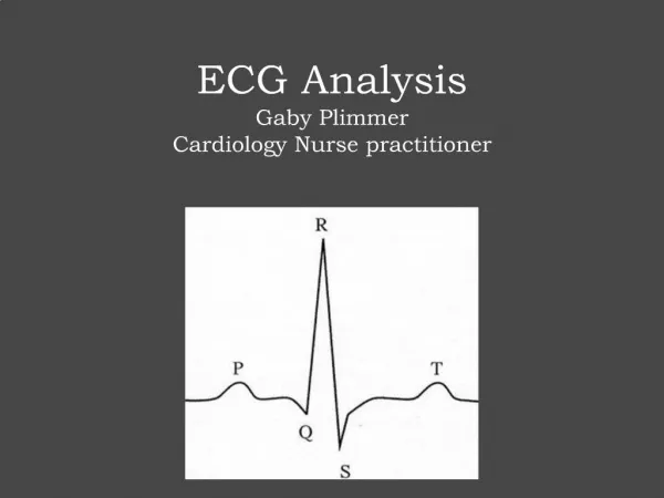
Where’s The “P” 2. Look at QRS3. Assess ST Segment
A 72-year-old man with a 35-year history of hypertension is evaluated because of dyspnea.
What is LVH? • Cornell: • R wave in aVL + S wave in V3 • >20 in ♀, >24 in ♂ • Voltage: • R wave in V5/V6 + S in V1 > 35mm • Non Voltage Changes: • LAA • LAD • IVCD • Asymmetric ST depression + T wave inversion
A 72-year-old man is noted to have bradycardia alternating with tachycardia during an orthopedic procedure.
ECG Diagnosis Atrial fibrillation (AF) is characterized by: 1. Rapid and irregular atrial fibrillatory waves at a rate of 350 to 600 imp/min 2. An irregularly irregular ventricular response of 90 up to 170 beats/min (Can be higher in some pts)
Pitfalls in Diagnosis 1. Fibrillatory waves may be inapparent on the standard and precordial leads. 2. Fibrillatory and U waves may have sufficient amplitude to look like P waves. 3. Extracardiac artifacts (eg, 60 cycle/min muscle tremors as in Parkinsonism) may simulate fibrillatory waves. 4. Regular R-R interval with AV dissociation or block with a lower junctional or ventricular pacemaker assumes control of the ventricles
A 65-year-old woman is evaluated because of new-onset atrial fibrillation and an embolic stroke. Echocardiography is performed. What does the echocardiogram show?
Spontaneous Echo Contrast • Increased RBC aggregation due to altered LAA flow dynamics and uncoordinated left atrial systole • Results in smoke-like echoes swirling in LA: SEC or “smoke” presumed to proceeding stage to thrombus formation • Seen in 50%- 65% of pts with AF
60-year-old woman with hypertension and diabetes mellitus is evaluated because of an 8-hour history of substernal chest pressure associated with dyspnea and diaphoresis.
MI - LOCATION • Anteroseptal: V1-3 • Inferior: II, III, F • Anterior: V2-4, or V1-V6 • Anterolateral: V4/5-V6, I, aVL • Right Ventricular: ST elevation V4R • Posterior: Tall R waves in V1, R/S ratio in V1>1
Normal ST elevation (concave) Early repolarization (concave) Normal variant with terminal T-wave inversion Marriott, NEJM, 2004.
LVH LBBB Brugada HyperK+ AS MI AS RBBB Acute Pericarditis Marriott, NEJM, 2004.
A 38-year-old woman is evaluated because of palpitations. She has a childhood history of the acute onset of rapid regular palpitations that she has learned to terminate with different vagal maneuvers. A 12-lead electrocardiogram taken during a period of palpitations is shown. What abnormality is seen?
Regular Sinus Tachycardia PSVT AVRT AVNRT SART Atrial Flutter PAT Irregular Atrial Fibrillation MAT Sinus Tachycardia with frequent PAC’s Atrial Flutter with variable AV block Commonly Encountered SVT
Most common SVT Two pathways within the AV node Rates: 120-250BPM Median age: 32+/-18 P waves often buried within QRS complex Inverted P waves in leads I, II, III and aVF Pseudo-r’ waves in V1 AVNRT
AVNRT <70ms
A 76-year-old woman with hypertension and paroxysmal atrial fibrillation is seen for a follow-up visit. Current medications are hydrochlorothiazide and digoxin.
Digitalis Toxicity • Classic combo disturbances: • Atrial tachycardia with AV block • Regular, accelerated junctional rhythm in AF
A 65-year-old man is evaluated because of lower-extremity edema and renal failure. Echocardiography is performed. What does the echocardiogram show?
A 75-year-old woman who was treated for heart block 3 years ago is seen for a follow-up visit.
An 87-year-old man with a pacemaker implanted for atrioventricular block is seen in follow-up.
An 80-year-old man underwent implantation of a single-chamber pacemaker for paroxysmal atrioventricular block 3 years ago. He now has a cerebrovascular accident.
VVI Mode • Responds to a sensed event. • Time value remaining in interval deleted • Output circuit disabled • Interval ends without a paced event • Commonly used for patients in chronic AF.
VVI Mode • Ventricular pacing • Ventricular pacing after ventricular escape interval(VV) • Ventricular sensed event, no pacing, VV reset • Ventricular pacing after reset ventricular escape interval
Atrial Flutter • Atrial tachycardia typically< 180 bpm • AT usually has isoelectric baseline • Atrial fluttter rates 240-200 bpm • Macroreentrant atrial rhythm with a reentry circuit involving large area of atrium • Commonly 2:1 AV conduction • Even ratios (2:1, 4:1) much more common than odd ratios (3:1, 5:1)
Type I or Typical atrial flutter Type II atrial flutter Atrial Flutter More negative in Lead II More positive F waves inferiorly M
A 59-year-old man is brought to the emergency department because of substernal chest pressure of 1 hour's duration.
Heart Block in MI • Inferior MI • Anterior MI:
A 53-year-old woman is evaluated because of pleuritic chest pain.
Classic Evolution in Acute Pericarditis • Stage 1: Concave ST segment elevation in almost all leads (no reciprocal ST depression) • Stage 2: ST segments decrease and T wave amplitude reduces • Stage 3: T waves invert • Stage 4: ECG normalizes
Acute Pericarditis • Look at lead aVR: • ST depression • PR elevation • Sinus Tachycardia • PR depression • Look for electrical alternans
An 84-year-old woman is hospitalized because of acute coronary syndrome.
A 74-year-old man with ischemic cardiomyopathy and an implanted cardioverter/defibrillator develops palpitations and presyncope.
VT- All favor • History: 90% of Ischemic CMP WCT=VT • Axis- “northwest/ right superior” • QRS Duration: • RBBB> 140ms • LBBB> 160ms • Precordial Concordance (esp negative) • AV Dissociation • Fusion beats • Capture beats
A 78-year-old woman is hospitalized because of pyelonephritis.
A 50-year-old man with severe ischemic cardiomyopathy is hospitalized because of syncope.
Bundle Branch Block QRS> 0.12ms • Left: • Broad monophasic R wave in leads I, V5, or V6 • Leads V1-2 reveal QS or rS pattern • Right: • Secondary r’ wave in V1 –o ften an M shape • Wide slurred S wave in I, V5, and V6
A 60-year-old man with hypertension and an evolving anterior wall myocardial infarction is evaluated in the emergency department.
Acquired: Polymorphic VT most commonly precipitated by long-short RR intervals
A 45-year-old male smoker is evaluated because of a 3-hour history of substernal chest pressure radiating to the left arm associated with nausea and vomiting.