Download
1 / 87
880 likes | 1.09k Vues
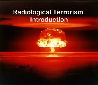
Medical Response to Nuclear and Radiological Terrorism. Stevan Cordas DO MPH Clinical Associate Professor TCOM/UNTHSC. Consultant Texas Department of Health - WMD Education Consultant American Osteopathic Association – Washington Bureau – WMD Certified Occupational Medicine (Toxicology)

E N D
Medical Response to Nuclear and Radiological Terrorism Stevan Cordas DO MPH Clinical Associate Professor TCOM/UNTHSC
Consultant Texas Department of Health - WMD Education • Consultant American Osteopathic Association – Washington Bureau – WMD • Certified Occupational Medicine (Toxicology) • Trained in Cleveland Institute of Nuclear Medicine • Former U.S. Army Medical Corps • Steering Committee - Medical Reserve Corps (Dallas, Tarrant, Denton and Collins County) • Author of WMD – AOA DO-online for CME
What Is Radiation? Radiation is energy transported in the form of particles or waves.
Exposure Vs. Contamination Exposure:irradiation of the body absorbed dose (Gray, rad) Contamination: radioactive material on patient (external)or within patient (internal)
Penetration Abilities of Different Types of Radiation Alpha Particles Stopped by a sheet of paper Radiation Source Beta Particles Stopped by a layer of clothing or less than an inch of a substance (e.g. plastic) Gamma Rays Stopped by inches to feet of concrete or less than an inch of lead Neutrons Stopped by a few feet of concrete CDC
Radiation Units Measure of Amount of radioactive material Ionization in air Absorbed energy per mass Absorbed dose weighted bytype of radiation Unit curie (Ci) roentgen (R) rad rem Quantity Activity Exposure Absorbed Dose Dose Equivalent
A Gray (Gy) The Gray (Gy) is a unit of absorbed dose and reflects an amount of energy deposited into a mass of tissue (1 Gy = 100 rads). In this lecture, the absorbed dose we are referring to is that dose inside the patient's body (i.e., the dose which is normally measured with personal dosimeters). For most purposes, one rem equal one rad. One mrem is one thousandth of one rem and is a common means of expressing radiation.
Radiation Doses andDose Limits Flight from Los Angeles to London 5 mrem Annual public dose limit 100 mrem Annual natural background 300 mrem Fetal dose limit 500 mrem Barium enema 870 mrem Annual radiation worker dose limit 5,000 mrem Heart catheterization (skin dose) 45,000 mrem Life saving actions guidance (NCRP-116) 50,000 mrem Mild acute radiation syndrome 200,000 mrem LD50/60 for humans (bone marrow dose) 350,000 mrem Radiation therapy (localized & fractionated) 6,000,000 mrem
Radioactive Material • Radioactive material consists of atoms with unstable nuclei • The atoms spontaneously change (decay) to more stable forms and emit radiation • A person who is contaminated has radioactive material on their skin or inside their body (e.g., inhalation, ingestion or wound contamination)
Half-life (HL) • Physical Half-Life Time (in minutes, hours, days or years) required for the activity of a radioactive material to decrease by one half due to radioactive decay • Biological Half-Life Time required for the body to eliminate half of the radioactive material (depends on the chemical form)
Effective Half-life The net effect of the combination of the physical & biological half-lives in removing the radioactive material from the body • Half-lives range from fractions of seconds to millions of years • 1 HL = 50% 2 HL = 25% 3 HL = 12.5%
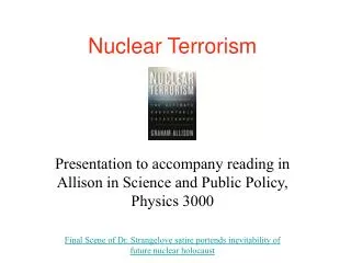
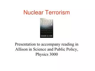
Potential Types of Weapons • Stolen nuclear material from a hospital, industry, university, power plant or disposal facility • Creation of a “dirty bomb” Generally thought to be the most likely scenario. • Nuclear detonation from a device. • Nuclear reactor sabotage
Hypothetical Suitcase Bomb CDC Chairman Dan Burton Committee – Demonstration of example “suitcase nuke” made from US nuclear shell
Examples of Radioactive Materials RadionuclideHalf-LifeActivityUse Cesium-137 30 yrs 1.5x10⁶ Ci Food Irradiator Cobalt-60 5 yrs 15,000 Ci Cancer Therapy Plutonium-23924,000 yrs 600 Ci Nuclear Weapon Iridium-192 74 days 100 Ci Industrial Radiography Hydrogen-3 12 yrs 12 Ci Exit Signs Strontium-90 29 yrs 0.1 Ci Eye Therapy Device
Examples of Radioactive Materials Iodine-131 8 days 0.015 Ci Nuclear Medicine Therapy Technetium-99m 6 hrs 0.025 Ci Diagnostic Imaging Americium-241 432 yrs 0.000005 Ci Smoke Detectors Radon-222 4 days 1 pCi/l Environmental Level
Types of Radiation Hazards • External Exposure - whole-body or partial-body (no radiation hazard to EMS staff) • Contaminated- • external radioactive material: on the skin • internal radioactive material: inhaled, swallowed, absorbed through skin or wounds Internal Contamination External Contamination External Exposure
Scope of Event Event Most Deaths Due to Number of Deaths Radiation None/Few Radiation Accident Few/Moderate Radioactive Blast Trauma (Depends on Dispersal size of explosion & Device proximity of persons) Blast Trauma Low Yield Large Thermal Burns (e.g. tens of thousands in NuclearWeapon an urban area even from Radiation Exposure 0.1 kT weapon) Fallout (Depends on Distance)
Facility Preparation • Activate hospital plan • Obtain radiation survey meters • Call for additional support: Staff from Nuclear Medicine, Radiation Oncology, Radiation Safety (Health Physics) • Plan for decontamination of uninjured persons • Establish triage area
Team Coordinator (leader) Emergency physician(s) Nurse (s) Triage Officer Administrator Radiation Safety Officer Maintenance Public Information Officer Security Laboratory Personnel Technical Recorder Develop Radiological Response Team
Consult With Radiation Experts • Radiation Safety Officer • Health Physicist • Medical Physicist • Conference of Radiation Control Program Directors (www.crcpd.org) CDC
Consult With Radiation Experts • Determining/documenting presence of radioactivity, activity levels, and radiation dose • Collecting samples to document contamination • Assisting in decontamination procedures • Disposing of radioactive waste
Detecting and Measuring Radiation • Instruments • Locate contamination - GM Survey Meter (Geiger counter) • Measure exposure rate - Ion Chamber • Personal Dosimeters - measure doses to staff • Radiation Badge - Film/TLD • Self reading dosimeter (analog & digital)
Biodosimetry Assessment Tool Armed Forces Radiobiology Research Institute www.afrri.usuhs.mil/
Facility Preparation • Plan to control contamination • Instruct staff to use universal precautions and double glove • Establish multiple receptacles for contaminated waste • Protect floor with covering if time allows
ED Staff Radiation Survey & Charting Contaminated Waste Radiation Survey STEP OFF PAD Waste Treatment Area Layout Separate Entrance CONTAMINATED AREA Trauma Room HOT LINE BUFFER ZONE Clean Gloves, Masks, Gowns, Booties CLEAN AREA
Immediate Medical Management • Triage • ARS • localized/ cutaneous • combined injury • Initial stabilization and treatment • Psychological effects • Record keeping/ Dose assessment
Protecting Staff from Contamination • Universal precautions • Survey hands and clothing with radiation meter • Replace gloves or clothing • that is contaminated • Keep the work area free of contamination • Key Points • Contamination is easy to detect and most of it can be removed • It is very unlikely that ED staff will receive large radiation doses from treating contaminated patients
Patient Management - Priorities Triage • Medical triage is the highest priority • Radiation exposure and contamination are secondary considerations • Degree of decontamination dictated by number of and capacity to treat other injured patients
Patient Management - Triage Triage based on: • Injuries • Signs and symptoms - nausea, vomiting, fatigue, diarrhea • History - Where were you when the bomb exploded or incident occurred? • Contamination survey
Psychological Casualties • Terrorist acts involving toxic agents (especially radiation) are perceived as very threatening • Mass casualty incidents caused by nuclear terrorism will create large numbers of worried people who may not be injured or contaminated
Psychological Casualties • Provide psychological support to patients and set up a center in the hospital for staff • Establish triage (monitoring and counseling) centers to prevent psychological casualties from overwhelming health care facilities • Staff counseling centers with physicians with a radiological background, health physicists with instrumentation and psychological counselors
Patient Management - Decontamination • Carefully remove and bag patient’s clothing and personal belongings (typically removes 95% of contamination) • Survey patient and, if practical, collect samples • Handle foreign objects with care until proven non-radioactive with survey meter
Patient Management - Decontamination • Decontamination priorities: • Decontaminate wounds first, then intact skin • Start with highest levels of contamination • Change outer gloves frequently to minimize spread of contamination
Patient Management - Decontamination (Cont.) • Protect non-contaminated wounds with waterproof dressings • Contaminated wounds: • Irrigate and gently scrub with surgical sponge • Extend wound debridement for removal of contamination only in extreme cases and upon expert advice • Avoid overly aggressive decontamination • Change dressings frequently
Patient Management - Decontamination (Cont.) • Decontaminate intact skin and hair by washing with soap & water • Remove stubborn contamination on hair by cutting with scissors or electric clippers • Promote sweating • Use survey meter to monitor progress of decontamination
Patient Management - Decontamination (Cont.) • Cease decontamination of skin and wounds • When the area is less than twice background, or • When there is no significant reduction between decon efforts, and • Before intact skin becomes abraded. • Contaminated thermal burns • Gently rinse. Washing may increase severity of injury. • Additional contamination will be removed when dressings are changed. • Do not delay surgery or other necessary medical procedures or exams…residual contamination can be controlled.
Patient Management - Patient Transfer Transport injured, contaminated patient into or from the ED: • Clean gurney covered with 2 sheets • Lift patient onto clean gurney • Wrap sheets over patient • Roll gurney into ED or out of treatment room
Facility Recovery • Remove waste from the Emergency Department and triage area • Survey facility for contamination • Decontaminate as necessary • Normal cleaning routines (mop, strip waxed floors) typically very effective • Periodically reassess contamination levels • Replace furniture, floor tiles, etc. that cannot be adequately decontaminated • Decontamination Goal: Less than twice normal background…higher levels may be acceptable
Injuries Associated With Radiological Incidents • Acute Radiation Syndrome (ARS) • Localized radiation injuries/ cutaneous radiation syndrome • Internal or external contamination • Combined radiation injuries with - Trauma - Burns • Fetal effects CDC
Radiation Sickness Acute Radiation Syndrome • Occurs only in patients who have received very high radiation doses (greater than approximately 100 rem or rads (1 Gy)) to most of the body • Dose ~ 15 rem • no symptoms, possible chromosomal aberrations • Dose ~ 50 rem • no symptoms, minor decreases in white cells and platelets
Acute Radiation Syndrome (Cont.)For Doses > 100 rem • Prodromal stage • nausea, vomiting, diarrhea and fatigue • higher doses produce more rapid onset and greater severity • Latent period (Interval) • patient appears to recover • decreases with increasing dose • Manifest Illness Stage • Hematopoietic • Gastrointestinal • CNS Time of Onset Severity of Effect
Acute Radiation Syndrome (ARS) • Radiation must be of penetrating type (X-rays, gamma rays or neutrons) • Most or all of body must be exposed. • The dose must be from an external source. • Dose must be delivered in a short time. Not fractionated.
The Three ARS Syndromes • Hematopoetic – Between 0.7 Gy and 10 Gy • Mortality rate is proportional to dosage. • Death from hemorrhage and infection • Absence of stem cells with leukopenia and thrombocytopenia. If they survive, anemia later.
Acute Radiation Syndrome (Cont.)Hematopoietic Component - latent period from weeks to days • Dose ~ 100 rem • ~10% exhibit nausea and vomiting within 48 hr • mildly depressed blood counts • Dose ~ 350 rem • ~90% exhibit nausea/vomiting within 12 hr, 10% exhibit diarrhea within 8 hr • severe bone marrow depression • ~50% mortality without supportive care