Download
1 / 56
640 likes | 821 Vues
Pathology is the study of diseases and mechanisms on the molecular or chemical level. This branch of medicine involves Clinical Pathology, Molecular Pathology, Hematopathology, Histopathology, Cytopathology, Blood Banking, and more. Learn about different divisions of pathology, techniques like FNAC and frozen sections, and the significance of histopathology in diagnosing diseases. Discover how Etiology, Pathogenesis, Morphology, and Clinical Significance play vital roles in understanding and studying diseases accurately.

E N D
Introduction to pathology Dr. Amitabha Basu MD
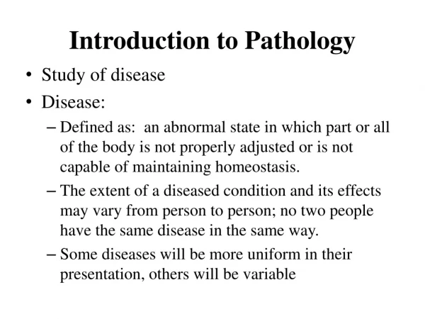
What is Pathology ? It is the study (logos) of the sufferings (pathos).
Basic Sciences P T H O L O G Y Clinical Medicine
Various Divisions of pathology • Clinical Pathology • Molecular Pathology • Hematopathology • Histopathology • Cytopathology • Blood banking
Clinical Pathology Automatic Analyzer
Clinical pathology • A branch of pathology concerned with patient care.
Molecular Pathology • A branch of pathology concerned with the study of the diseases and mechanisms of disease on a molecular or chemical level.
Human immunodeficiency virus, viral particles at medium magnification adjacent to cell surface, electron micrograph.
Hematopathology • This branch deal with the Disease of the Blood. This peripheral blood smear is stained with the Wright's stain. (Leishman stain)
CBC (complete blood count) : from Coulter blood cell counter
Cytopathology: Study of cellular change in the diseased tissue.
Cytopathology Study of cell Tissue composed of many cell of similar function
Two methods to obtain the cells • Exfoliative cytology ( collect and examine the cell that falls –off from the tissue) • FNAC (Fine needle Aspiration Cytology): cell obtained with a fine needle.
What is that mean ? • Exfoliated cells ( eg from Cervix) for quick diagnosis of malignancy • Stain Uses: • Papanicolaou Stain Pap Smear
Cervical Cytology • It helps in early diagnosis of cancer of Uterine Cervix. So, you can prevent a cancer like this!!
FNAC [ fine needle aspiration cytology ] Cells obtained from an abnormal mass in the body : EG Breast lump
Aspirated Thyroid cells shows features of malignancy : Aspirated cells stained with Giemsa Stain It is quick. Less expensive No hospital stay required
Histopathology Study of tissue Tissue composed of many cell of similar function
Histopathology- a technique to identify a disease by looking at the tissue! House of final diagnosis. • Tissue is collected and fixed with formalin- overnight- it takes time!. • It is then embedded in paraffin and cut with microtome- 3 micron thick. • This thin tissue is then stained with Hematoxiline & eosin( H&E) stain. • Following that it is mounted with DPX and cover slip.
Normal Squamous cell and Histopathology of Squamous cell carcinoma. Definition of histopathology: Pathological Study of the minute structure, composition, and function of diseased tissues.
Histopathology An important procedure to rule out or confirm malignancy. Stain Used : Hematoxylin and Eosin Stain [ H & E ]
Biopsy • The removal and examination of a sample of tissue from a living body for diagnostic purposes
Biopsy sample then sent to the Histopathology laboratory Tissue were kept in the Formalin for Fixation and to avoid autolysis
Frozen section • Paraffin section takes time--- • If you need a quick section…..we harden the tissue by freezing it…frozen section.
Frozen Sections • It is necessary to get a rapid diagnosis of a pathologic process. • The piece's are snap frozen in a cold liquid or cold environment (-20 to -70 Celsius). • Freezing makes the tissue solid enough to section with a microtome.
Use of frozen section technique. • To check to presence of tumor in surgical resected ends, while removing a tumor from the body. • To check for the presence of metastasis tumor in lymph node. • To identify fat.
This is not enough! So we need special stains!
Blood Bank • Optimal Blood Testing, Preservation and Utilization of Blood and blood products.
Welcome once again to the world of Pathology Why ? How ? Where ? = Answer this and you will get your diagnosis
How to study pathology ? Easy • Learn • Whyis the disease = Etiology • What are the types = Classification • How the disease occur = Pathogenesis • Where = Morphological Change of the organ effected • What happens then = Clinical significance. Follow this pattern and you will never forget pathology
Understanding of a few terms: and also study pathology in this sequence. • Etiology • Pathogenesis • Morphology • Gross change of a diseased organ/tissue • Microscopical change of the tissue and cells. • Functional Derangement and Clinical Significance
Etiology • Cause of the disease. • Example : Chronic Alcoholism is the etiology of fatty liver.
Pathogenesis Definition : Mechanism of disease formation • Alcohol produce injury to the liver cells , following that Liver cells (Hepatocytes) become unable to metabolize Fatty acid. • And it accumulate in the liver cells to produce fatty liver.
Pathogenesis : narrowing of the coronary artery : Myocardial Infarction.
Gross visible change of a diseased organ/tissue. Microscopical change of the tissue and cells. Morphology : 2 parts