Download
1 / 38
460 likes | 1.29k Vues
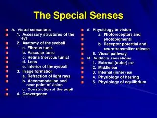
A. Visual sensations 1. Accessory structures of the eye 2. Anatomy of the eyeball a. Fibrous tunic b. Vascular tunic c. Retina (nervous tunic) d. Lens e. Interior of the eyeball 3. Image formation

E N D
A. Visual sensations 1. Accessory structures of the eye 2. Anatomy of the eyeball a. Fibrous tunic b. Vascular tunic c. Retina (nervous tunic) d. Lens e. Interior of the eyeball 3. Image formation a. Refraction of light rays b. Accommodation and near point of vision c. Constriction of the pupil 4. Convergence 5. Physiology of vision a. Photoreceptors and photopigments b. Receptor potential and neurotransmitter release 6. Visual pathway B. Auditory sensations 1. External (outer) ear 2. Middle ear 3. Internal (inner) ear 4. Physiology of hearing 5. Physiology of equilibrium The Special Senses
Surface Anatomy and Accessory Structures of the Eye 1. eyelids 2. eyelashes 3. eyebrows 4. extrinsic eye muscles 5. lacrimal apparatus
Lacrimal Apparatus • 1. lacrimal gland • 2. lacrimal ducts • 3. lacrimal puncta • 4. lacrimal canals • 5. nasolacrimal duct
Anatomy of the Eyeball • 1. fibrous tunic a. cornea b. sclera _______________ • canals of Schlemm Sclera pupil iris
Anatomy of the Eyeball Con’t • 2. vascular tunic a. choroid b. ciliary body (1) ciliary muscle (2) ciliary processes c. iris (1) sphinctor pupillae (2) dilator pupillae Dilation Constriction Dilator pupillae Sphinctor pupillae
Anatomy of the Eyeball Con’t • 3. nervous tunic (retina) a. pigmented layer b. nervous layer Terms: fovea centralis optic disc (blind spot)
Anatomy of the Eyeball, Con’t • 3. b. nervous layer (1) photoreceptor layer (a) rods (b) cones (2) bipolar layer (a) bipolar cells (b) amacrine cells (c) horizontal cells (3) ganglion cell layer
Lens • 1. biconvex • 2. crystallins • 3. suspensory ligaments • 4. focuses light Canals of Schlemm Ciliary body Ciliary Processes iris Suspensory ligaments lens
Cavities of the Eye • 1. anterior cavity a. posterior chamber b. anterior chamber c. aqueous humor d. fluid flow e. canals of Schlemm f. intraocular pressure 2. posterior cavity a. vitreous humor b. tugor c. not replaced Posterior Chamber Anterior Chamber
The formation of images on the retina involves three processes: • 1. refraction of light rays • 2. accommodation of the lens • 3. constriction of the pupil
Refraction of Light 1. light passes through different media 2. light bends at interfaces 3. 60% at cornea 4. 40% by lens
Accomodation • 1. biconvex lens • 2. point of intersection • 3. near object = more convex • 4. far object = less convex • 5. role of ciliary muscle
Constriction of the Pupil • 1. part of accommodation reflex • 2. limits peripheral light
Convergence of the Eyes • 1. single binocular vision • 2. corresponding points • 3. complementary photoreceptors • 4. near vision = medial rotation of eyes
Photoreceptors • 1. central fovea • 2. visual axis • 3. rods and cones • 4. outer segments • 5. visual pigments • 6. glutamate • 7. dark vs light conditions
Visual Acuity Rod cells not present in central fovea increase in concentration to ora serrata dim light and peripheral vision 6 – 600 rod cells converge on 1 bipolar cell, as well as amacrine and horizontal cells. Cone cells 100% cone cells in central fovea diminish in number toward ora serrata three different types: blue, red, green cone cells bright light required to break pigments 1:1 relationship with bipolar cells visual acuity and color vision
DARK VERSUS LIGHT CONDITIONS X DARK rod cell Na+ channels open visual pathway started membrane depolarizes Na+ inflow stimulates glutamate release glutamate inhibits bipolar cells LIGHT rod cell Na+ channels close membrane hyperpolarizes no Na+ inflow prevents glutamate release bipolar cells initiate action potential visual pathway started
The Visual Pathway • The visual pathway begins with stimulation of the bipolar neurons by photoreceptor cells. • 1. 6 - 600 rod cells converge on 1 bipolar cell, as well as amacrine and horizontal cells • 2. Cone cells have a 1:1 relationship with bipolar cells
Visual Pathway • 1. bipolar cells • 2. ganglion cells • 3. optic nerve (II) • 4. optic chiasma a. medial retina vs b. lateral retina • 5. optic tracts • 6. thalamus • 7. optic radiations • 8. primary visual areas
External Ear • Auricle (pinna) • External auditory meatus • Tympanic membrane • Ceruminous glands
Middle Ear • 1. petrous portion of temporal bone • 2. mastoid air cells • 3. auditory tube • 4. ossicles ________________ • 5. oval window • 6. round window
Auditory Ossicles • 1. malleus • 2. incus • 3. stapes ____________ • stapedius • tensor tympani
Internal Ear • 1. bony labyrinth a. 3 semicircular canals (1) frontal (2) horizontal (3) sagittal b. vestibule c. cochlea ________________ perilymph
Internal Ear • 1. bony labyrinth a. 3 semicircular canals (1) frontal (2) horizontal (3) sagittal b. vestibule c. cochlea ________________ perilymph
Internal Ear • 2. membranous labyrinth a. 3 semicircular ducts b. utricle and saccule c. cochlear duct _________________ endolymph
Cochlea • 1. scala vestibuli • 2. scala tympani • 3. scala media (cochlear duct)
Cochlea • 1. scala vestibuli • 2. scala tympani • 3. scala media (cochlear duct) • 4. vestibular membrane • 5. basilar membrane • 6. spiral organ of Corti • 7. 16,000 hair cells • 8. tectorial membrane
Physiology of Equilibrium • 1. static equilibrium (saccule and utricle) perception of head orientation when the body is stationary • 2. dynamic equilibrium (mainly semicircular ducts) perception of motion or acceleration (linear vs angular) *linear acceleration-utricle and saccule
Static Equilibrium • 1. utricle and saccule • 2. hair cells • 3. otoliths of macula • 4. inertia
Dynamic Equilibrium • 1. semicircular ducts • 2. hair cells • 3. ampullae • 4. cupula • 5. inertia