Download
1 / 33
330 likes | 362 Vues
Discover the foundation of genetics in the 1920s, including chromosomes, DNA structure, amino acids, and transformative experiments. Explore the pivotal discoveries by Griffith, Avery, Hershey, Chase, and Watson-Crick. Unlock the secrets of DNA's double helix and understand the chemistry behind base pairing. Learn about Chargaff's rules and Franklin's X-ray diffraction data.

E N D
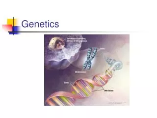
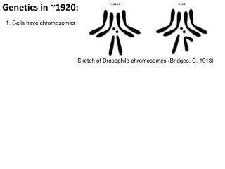
Genetics in ~1920: 1. Cells have chromosomes Sketch of Drosophila chromosomes (Bridges, C. 1913)
Genetics in ~1920: 1. Cells have chromosomes Sketch of Drosophila chromosomes (Bridges, C. 1913) 2. Specific locations on chromosomes control different phenotypes Mutant phenotypes Short aristae Cinnabar eyes Vestigial wings Brown eyes Black body 0 48.5 57.5 67.0 104.5
Genetics in ~1920: Chromosomes are made of: DNA Protein Which one is the genetic material?
DNA: consists of nucleotides 5’ 4’ 1’ 3’ 2’ The structure of a nucleotide
DNA: consists of nucleotides (4 types) 5’ 4’ 1’ Pyrimidines 3’ 2’ The structure of a nucleotide Purines
Nitrogenous bases Sugar–phosphate backbone 5 end DNA is a polymer -Nucleotides are joined together by linking 5’ and 3’ carbons of sugar groups to phosphate groups - A chain of nucleotides has a 5’ end and a 3’ end Thymine (T) Adenine (A) Cytosine (C) DNA nucleotide Phosphate Sugar (deoxyribose) 3 end Guanine (G)
Protein: Consists of amino acids Carboxyl group Amino group
Nonpolar Protein: Consists of amino acids (20 different types) Glycine (Gly or G) Valine (Val or V) Leucine (Leu or L) Isoleucine (Ile or I) Alanine (Ala or A) Trypotphan (Trp or W) Methionine (Met or M) Phenylalanine (Phe or F) Proline (Pro or P) Polar Glutamine (Gln or Q) Serine (Ser or S) Threonine (Thr or T) Cysteine (Cys or C) Tyrosine (Tyr or Y) Asparagine (Asn or N) Electrically charged Acidic Basic Carboxyl group Amino group Glutamic acid (Glu or E) Histidine (His or H) Aspartic acid (Asp or D) Lysine (Lys or K) Arginine (Arg or R)
Peptide bond A polypeptide (a) Fig. 5-18 Side chains Peptide bond Backbone Amino end (N-terminus) Carboxyl end (C-terminus) (b)
Griffith (1928) Transformation of bacteria Mixture of heat-killed S cells and living R cells EXPERIMENT Living R cells (control) Living S cells (control) Heat-killed S cells (control) RESULTS Mouse dies Mouse healthy Mouse healthy Mouse dies Living S cells
Griffith (1928) Transformation of bacteria Mixture of heat-killed S cells and living R cells EXPERIMENT Living R cells (control) Living S cells (control) Heat-killed S cells (control) RESULTS Mouse dies Mouse healthy Mouse healthy Mouse dies Living S cells
Avery (1944) DNA is the transforming material How could he have shown this?
Hershey + Chase (1952) T4 infection of E. coli Phage head Tail sheath Tail fiber DNA 100 nm Bacterial cell
Hershey + Chase (1952) T4 infection of E. coli EXPERIMENT Radioactive protein Phage Bacterial cell DNA Batch 1: radioactive sulfur (35S) Radioactive DNA Batch 2: radioactive phosphorus (32P)
Hershey + Chase (1952) T4 infection of E. coli EXPERIMENT Empty protein shell Radioactive protein Phage Bacterial cell DNA Batch 1: radioactive sulfur (35S) Phage DNA Radioactive DNA Batch 2: radioactive phosphorus (32P)
Hershey + Chase (1952) T4 infection of E. coli EXPERIMENT Empty protein shell Radioactivity (phage protein) in liquid Radioactive protein Phage Bacterial cell DNA Batch 1: radioactive sulfur (35S) Phage DNA Centrifuge Pellet (bacterial cells and contents) Radioactive DNA Batch 2: radioactive phosphorus (32P) Centrifuge Radioactivity (phage DNA) in pellet Pellet
Hershey + Chase (1952) T4 infection of E. coli EXPERIMENT Empty protein shell Radioactivity (phage protein) in liquid Radioactive protein Phage Bacterial cell DNA Batch 1: radioactive sulfur (35S) Phage DNA Centrifuge Pellet (bacterial cells and contents) Radioactive DNA Batch 2: radioactive phosphorus (32P) Centrifuge Radioactivity (phage DNA) in pellet Pellet
Chargaff (1949) 02_UnTable01.jpg
1953: The double helix Watson and Crick Rosalind Franklin
Key aspects of the Watson-Crick model 5 end - The 2 strands are in shape of a double helix -10.5 base pairs per turn of the helix Hydrogen bond 3 end 1 nm 3.4 nm 3 end 0.34 nm 5 end (a) Key features of DNA structure (b) Partial chemical structure
Nitrogenous bases Sugar–phosphate backbone 5 end Data used to deduce double helix: • Chemical structure of DNA polymer • Chargaff’s rules • Franklin’s X-ray diffraction data Thymine (T) Adenine (A) Cytosine (C) DNA nucleotide Phosphate Sugar (deoxyribose) 3 end Guanine (G)
(b) Franklin’s X-ray diffraction photograph of DNA • This told them: • 2 anti-parallel DNA strands • Helical shape • Width, period of helix
Key aspects of the Watson-Crick model 5 end -2 anti-parallel strands of DNA -Sugar-phosphate backbone on outside, bases on inside -Bases form pairs through hydrogen bonding 3 end Hydrogen bond 1 nm 3.4 nm 3 end 0.34 nm 5 end (a) Key features of DNA structure (b) Partial chemical structure
Purine Pyrimidine Base pairing Adenine (A) Thymine (T) Cytosine (C) Guanine (G)
Purine + purine: too wide Fig. 16-UN1 Pyrimidine + pyrimidine: too narrow Purine + pyrimidine: width consistent with X-ray data
Watson and Crick- 1953 “It has not escaped our notice that the specific pairing we have postulated immediately suggests a possible copying mechanism for the genetic material.”
A T T A C G G C Fig. 16-9-2 A T A T T A T A C C G G (b) Separation of strands (a) Parent molecule
A T A T A T A T C G C G C G C G Fig. 16-9-3 A T A T A A T T T A T A T T A A C C G C G C G G (c) “Daughter” DNA molecules, each consisting of one parental strand and one new strand (b) Separation of strands (a) Parent molecule
First replication Second replication Parent cell (a) Conservative model Fig. 16-10 (b) Semiconserva- tive model (c) Dispersive model
EXPERIMENT Bacteria cultured in medium containing 15N Bacteria transferred to medium containing 14N 2 1 Fig. 16-11a RESULTS DNA sample centrifuged after 20 min (after first application) DNA sample centrifuged after 20 min (after second replication) Less dense 3 4 More dense
CONCLUSION First replication Second replication Conservative model Fig. 16-11b Semiconservative model Dispersive model