lips
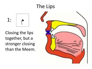
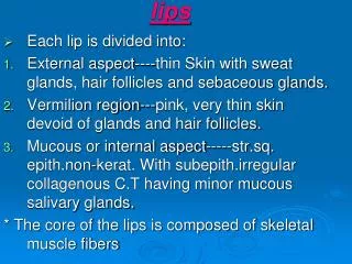
lips. Each lip is divided into: External aspect----thin Skin with sweat glands, hair follicles and sebaceous glands. Vermilion region---pink, very thin skin devoid of glands and hair follicles.

lips
E N D
Presentation Transcript
lips • Each lip is divided into: • External aspect----thin Skin with sweat glands, hair follicles and sebaceous glands. • Vermilion region---pink, very thin skin devoid of glands and hair follicles. • Mucous or internal aspect-----str.sq. epith.non-kerat. With subepith.irregular collagenous C.T having minor mucous salivary glands. * The core of the lips is composed of skeletal muscle fibers
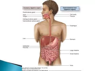
Oral cavity. Oral mucosa: *Gingiva, dorsal surface of the anterior two thirds of the tongue, and hard palate are covered by keratinized or partially (parakeratinized) str.sq.epith. with underlying dense irregular collagenous C.T. *The reminder of oral cavity is covered by non-keratinized str. Sq. epith. With underlying looser collagenous C.T.
Teeth • Each tooth is suspended in bony socket, the alveolus by the periodontal (dense irregular collagenous C.T.) • Each tooth is formed of: 1-Crown—visible part. 2-Root----in the alveolus. 3-Cervix---inbetween. *The inner part of tooth is the pulp that contain soft vascular C.T contains bl. and lymph Vessels and nerves
Mineralized components • They are : • a.Dentin----surrounds the pulp and is covered with; • b.Enamel–that cover the crown. • c.Cementum----that cover the root.
Palate It is composed of: • Hard palate (keratinized-st. sq.epith.) • Soft palate (non-ker.st.sq.epit.) • Uvula (non-ker.st.sq.epith) They separate the nasal cavity from the oral one.
Tongue • It has: 1-Dorsal surface---its ant. two thirds is covered with ker. St. sq.epith. and separated from post one third (covered with non-ker. St. sq.epith) by a shallow, V-shaped groove, the sulcusterminalis. 2-Ventral surface---non-ker. St.sq.epith. 3-Core of skeletal muscle fibers. *The dorsal surface of posterior one third has lingual tonsils.
Lingual papillae They are located on the dorsal & lateral aspect of the tongue. There are four types: 1.Filiform papilla : slender structure, covered by ker.st. sq. epith.DO NOT have taste buds. 2.Fugiform papilla---as mushroom has slender stalk connects a broad cap to tongue surface.It is covered by non-ker. Str. Epith.It has taste buds on dorsal surface of the cap.
3.Foliate papillae: are located along the posterior aspect of the tongue. They have taste buds in neonate only. They have furrows in which glands of Von Ebner (serous) open. 4.Circumvallate papillae---8 to 12 just ant. To sulcus terminalis. They have Von Ebner serous glands. They have taste buds on their sides only.
Taste buds • Are intra-epithelial sensory organs for perception of taste. • Each taste bud is formed of: 1.Dark cells (type I) 2.Light cells (type II) 3.Intermediat cells (type III) 4.Basal cells (type IV) *Nerve fibers synapse with types I, II, and III (they have long microvilli protruding from taste pores)
Salivary glands There two types of salivary glands: Minor salivary glands (scattered in the mucosa of oral cavity-mucous secreting). Major salivary glands ( Parotid, sub mandibular and sub lingual)
Major salivary glands • There are three pairs of major salivary glands, Parotid, submandibular, and sublingual. • They have C.T capsule. provides septa that divide the glands into lobes and lobules (Stroma). • Their parenchyma consists of secretory portion (tubuloalveolar glands) and ducts portion.
Secretory portions • Are formed of serous and/or mucous serous secretory cells arranged as acini (alveoli-serous) or tubules (mucous) that are couched by myoepithelial cells. • Myoepithelial (basket )cells. They share the basal lamina of acinar cells (hemidesmosomes). They envelope the cells of secretory acinus and intercalated ducts (desmosomes) They have several long processes. They are rich in actin and myosin. They press on the acinus to release the product.
Serous cells • Secrete proteins and polysaccharides. • Are pyramidal with single round, basally located nuclei. • Are rich in rER, Golgi complex, basal mitochondria, and epically situated secretory granules. • They have tight junctions, intercellular canaliculi and interdigitated baso-lateral processes.
Mucous cells • Are short pyramidal cells with basal flattened nuclei. • Have few mitochondria, rER, but rich in Golgi complex (to form carbohydrates). • Have less lateral processes and intercellular canaliculi than serous cells. • Apices of cells are rich in secretory granules.
Duct portions • Are highly branched ducts. • Begin with the smallest intercalated ducts that formed of small cuboidal cells having myoepithelial cells. • Intercalated ducts merge to form striated ducts which are cuboidal to columnar cells with basolateral folds containing mitochondria. They join together to form intralobular ducts that unit to form interlobular ducts that join to form intralobar and interlobar ducts. • Terminal ducts open into the oral cavity.
Parotid Gland • The largest salivary gland but produce 30% of salivary output. • It secrets pure serous secretion that rich in amylase enzyme, lactoferrins, lysozymes and secretory IgA
Submandibular Gland • It produces 60% of salivary output. • It is mixed but the major portion (90%) is serous and 10% is mucous. • It has few serous demilunes that capped the mucous tubular secretory unit.
Sublingual Gland • It is very small and responsible for 5% of salivary secretion. • It is composed of mucous tubules with serous demilunes. • It produces mixed , but mostly mucous saliva. • Its duct system does not form terminal duct, instead several ducts open into the floor of oral cavity.
Alimentary Canal • Is the tubular portion of digestive system. • About 9 meters and subdivided into: esophagus, stomach, small intestine (duodenum, jejunum and ileum), and large intestine (cecum, colon, rectum, anal canal, and appendix)
General structure of Alimentary tract It is formed of 4 concentric layers: I.MucosaII.SubmucosaIII.Muscularisexterna IV.Serosa (adventitia) I-MUCOSA It is formed of : 1-Epithelium. 2-Lamina propria (Vascular C.T) that contains lymph vessels, nodules and glands. 3-Muscularis mucosa:longitudinal smooth muscle.
II-SUBMUCOSA • Is formed of dense irregular elastic C.T. • It has glands ONLY in esophagus and duodenum. • It is rich in blood and lymph vessels. • It has enteric nervous plexus (Meissner”s plexus) that houses also post ganglionic parasympathetic nerve cell bodies.
III-MUSCULRIS EXTERNA • It is responsible for peristaltic activity. • It is composed of smooth muscle (EXCEPT in esophagus, has both smooth & skeletal fibers). • They are arranged helically. • Usually organized as inner circular and outer longitudinal. • Between the two layers they have Auerbach’s myenteric plexus. • that houses also post ganglionic parasympathetic nerve cell bodies
Musculsris externa Auerbach’s myenteric plexus
IV-SEROSA OR ADVENTITIA • It covers the muscularis externa. • It is formed of thin layer of vascular C.T. • If it is surrounded by simple squamous epith.of the visceral layer of peritoneum (mesothelium)---It is called serosa. • If the organ is retropritoneal i.e NO epith.—It is called adventitia.
Esophagus Mucosa---stratified sq.epith non-ker,fibroelasic lamina propria and longitudinal muscularis mucosa (smooth muscle). L.P has cardiac glands (mucous) near the pharynx (upper region) and stomach (lower region). Submucosa– has mucous esophageal glands proper. Muscularis externa of upper third is skeletal muscle, middle third is both smooth&skeletal and the lowest third is smooth muscle. Adventitia until pierces diaphragm---serosa
Mucosa and submucosa of esophagus submucosa
Gastro-oesophageal junction esophagus stomach
Fundic mucosa Fundic glands have: Short pits—one forth of mucosa. Simple or branched tubular glands. Are rich in parietal & chief cells.
Surface columnar gastric cell They secrete thick mucous
Parietal cell Acidophilic cells, apical invaginations that have canaliculi lined by microvilli. Secrete HCl and gastric intrinsic factor (absorption of Vit.B12 in the ileum).
Chief cell *Basophilic cytoplasm. *Secrete pepsinogen, rennin and gastric lipase
Fundic glands • Their pits are short about one quarter of length of mucosa. • Are simple or branched tubular glands. • Are numerous and crowded. Pyloric glands * Their pits are deep---about half the length of mucosa. * They are branched and convoluted---many cross sections. * The predominant cells are mucous neck cells that secrete both mucous and lysozyme.
Small intestine • It has 3 regions: duodenum, jejunum and ileum. • It has: * Plicae circulares---transverse folds of mucosa and submucosa. *Villi—Finger like protrusions of lamina propria that is covered with epithelium.They contain Bl. &lymphatic vessels (lacteal), smooth muscle fibers, loose CT and lymphoid cells. * Micrvilli—modifications of the apical region of plasmalemma of epithelial cells of villi and surface epithelium. Crypts of Lieberkuhn-invaginations of epithelium into the L.P between the villi form glands