Download
1 / 21
210 likes | 319 Vues
Announcements, Feb. 12. Happy Darwin Day! (b. Feb. 12, 1809) Reading for today: 172-186 on membrane proteins. Reading for Wednesday: 191-207 on membrane transport. SDS-PAGE homework problem due

E N D
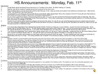
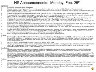
Announcements, Feb. 12 • Happy Darwin Day! (b. Feb. 12, 1809) • Reading for today: 172-186 on membrane proteins. • Reading for Wednesday: 191-207 on membrane transport. • SDS-PAGE homework problem due • Also on Wednesday: Representatives in biology will be at the Annual Summer Job Fair, UC Rotunda and Terrace Rooms, from 11 AM to 4 PM. • Reading for Friday: 207-216 on energetics of membrane transport. • Electron spin resonance (ESR) spectroscopy measures lipid mobility in membranes • Similar to NMR, labeling with nitroxyl group (>N-O) • Atomic force microscopy measures heights of various parts of specimen at the molecular level.
Suggestions for studying from your fellow students (>90% on Exam 1) • “I went to the study session, had a study group, and read over the powerpoints.” • “I read through the powerpoint slides.” • “I started studying 5 days before & studied 2 sets of notes each night & then all on the Thursday night before the test. I also looked at diagrams on the power points.” • “I rewrote my notes and made notes from that on the stuff I didn’t know as well. Then studied mainly that material.” • “… Re-wrote notes, made up questions and answered.”
Membrane Proteins SDS-PAGE Membrane proteins in red blood cells Classes of MB proteins Orientation and glycosylation of MB proteins After reading the text, attending lecture, and reviewing lecture notes, you should be able to Explain how proteins can be separated by SDS-PAGE, and apply your knowledge to solve SDS-PAGE problems. Describe the classes of membrane proteins and how they can be removed from membranes. Outline/Learning Objectives
SDS-PAGE Reagents • Sodium dodecyl sulfate • Strong ionic detergent • Removes proteins from MB, solubilizes hydrophobic amino acids • Unfolds and coats proteins w/ negative charge • Triton X-100 • Mild non-ionic detergent • Removes proteins from MB • Solubilizes proteins but does not unfold them.
SDS-PAGE Reagents • Polyacrylamide forms gel matrix • small proteins go through fast • large proteins go through slowly • -mercaptoethanol breaks S-S bonds • with: reducing conditions break subunits apart • without: non-reducing conditions keep subunits together • Allows determination of number of subunits HS-CH2-CH2-OH
Functions of MB Proteins • Receptors or signals, e.g. glycophorin • Structural, e.g. spectrin, ankyrin, Band 4.1 • Transporters, e.g. glucose transporter, Band 3 • Channels, e.g. Na channels in excitable cells • Enzymes, e.g. G3PDH • Electron transport proteins in mitochondria, chloroplasts • Intercellular adhesion and communication, e.g. gap junctions
Evidence for mosaic of proteins: Freeze-fracture SEM
Freeze-Fracture SEM of membranes < Artificial bilayer w/o protein Artificial bilyaer w/protein >
C. Classes of Membrane Proteins transmembrane 1. Integral membrane proteins: require detergent to remove from MB 2. Peripheral membrane proteins: removed by milder treatments 3. Lipid-anchored membrane proteins: in lipid rafts
Solubilization of integral membrane protein by nonionic detergent Critical micelle concentration
SDS-PAGE Problem • You are given a preparation of kangaroo membranes (M), part of which looks like this (assume membrane is sealed): • You do the following experiment, where an arrow indicates centrifugation: • You also treat M with protease, on the side of the bilayer indicated in the diagram. This sample is called PRO. • All samples (M, S1, P1, S2, P2, PRO) are mixed with SDS and run on a denaturing polyacrylamide gel. Diagram what you expect to see in the gel for each sample. Protease Protein B Out In membrane Protein A Isolated membranes (M) Sup S1 Pel P1 Add non-ionic detergent (to solubilize membranes) Sup S2 Pel P2 Salt wash Spin Spin
M S1 P1 S2 P2 PRO M S1 P1 S2 P2 PRO Solution and Homework • Part 1: • Part 2: You then get adventurous, and look at the membrane from an aardvark cell, repeating the exact same protocols as for the kangaroo membranes. You also run a gel, as above, and see this: • Homework: Draw a diagram of what the aardvark membrane looks like (Hint: the protein may cross the membrane more than once).
1. Integral Membrane Proteins Example of connexin: 4 positive peaks from hydropathy analysis predicts the protein has 4 transmembrane domains. • Strong treatments (detergents) are required to remove from MB. • Amphipathic molecules • Transmembrane regions are -helical with hydrophobic R groups facing out - usually 20-30 amino acids.
2. Peripheral and 3. Lipid-Anchored Membrane proteins Peripheral MB proteins • Weak treatments (change in pH or ionic strength, removal of Ca2+) remove from MB since bound by electrostatic interactions or H-bonds. • Can be on outside or inside: spectrin, ankyrin, and Band 4.1 are inside examples from RBCs. Lipid-anchored MB proteins • Covalently bound to membrane lipids. • Most bound to fatty acids on inner leaflet. • Some bound to outer leaflet linked to GPI (a glycolipid in external monolayer) • May be enriched in lipid rafts
Glycophorin and Bacteriorhodopsin • Bacteriorhodopsin was one of first membrane proteins whose 3D structure was determined. • It functions as a light-driven proton pump
D. Many membrane proteins are glycosylated • In addition to lipids and proteins, most membranes have significant amounts of carbohydrates • Erythrocyte - 52% protein, 40% lipid, 8% carb. • Glycolipids account for only small portion of membrane carbohydrates; most is in form of glycoproteins. • Addition of carbohydrate side chain to a protein is glycosylation.
N-linked and O-linked glycosylation • Linkage to either N or O on R groups • Function of glycoproteins: usually in plasma membranes, role in cell-cell recognition along with membrane receptors • Glycosylation occurs in ER and Golgi
Purpose of protein glycosylation • Synthesis of complex carbohydrates requires a separate enzyme for each different step, unlike other polymerization reactions. • May be several functions, not well-understood • Presence of oligosaccharides makes glycoprotein more resistant to digestion by extracellular proteases. • Glycosylation also may be important for receptor-ligand binding. • CHO-binding proteins are called lectins