Download
1 / 37
580 likes | 1.68k Vues
Chapter 10. Ischemia-reperfusion injury (IRI). Yuxia Zhang. Department of Pathophysiology, Anhui Medical University. Contents. Concepts: IRI, oxygen/calcium/pH paradox Causes and conditions of IRI Mechanisms of IRI injury Metabolic and functional alterations

E N D
Chapter 10 Ischemia-reperfusion injury (IRI) Yuxia Zhang Department of Pathophysiology, Anhui Medical University
Contents • Concepts: IRI, oxygen/calcium/pH paradox • Causes and conditions of IRI • Mechanisms of IRI injury • Metabolic and functional alterations • Prevention and treatment principle
Introduction • at 1960,Jenning:MI/R I • 1968, brain;1972, kindey;1978, lung;1981, intestinal;and so on • Clinical phenomenon: bypass surgery,shock treatment,organ transplantation, thrombolysis, recovery of hearts after ischemic arrest,Percutanueous Transluminal Coronary Angioplasty (PTCA) • I/R I is a common phenomenon • paradoxical phenomenon
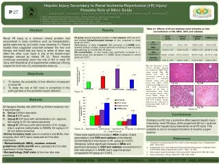
1.Concept • IRI the reestablishment of blood flow after prolonged ischemia aggravates the tissue damage.
pH paradox ischemiaacidosis , disorder of function andmetbolism on cell severe IRIpH paradox • calcium paradox pre-perfuse rat heart with no calciumperfusion for 2minperfuse calcium perfusion, cell release enzyme myofibril over-constract, electron signals abnormal, calcium paradox
Oxygen paradox Hypoxia liquid perfuse organ or culture without oxygen injury restore perfusionsevere injury
2. Cause of ischemia-reperfusion injury and affecting factor Recover from cardiac arrest Organ transplantation Lysing thrombi (1)cause
(2)Affecting factor 5-10min: arrhythmia small animals 20-30min: ventricular tremor • Duration of ischemia 20-40min: reversible injury big animals 40-60min:irreversibleinjury diversity between small and big animal
Branch circulation:chronic • O2 consumption rate T, pressure,pH,Na+,Ca2+ protection • condition of • reperfusion [K+ ], [Mg2+] damage T, pressure,Na+,Ca2+
3. Mechanisms of IRI • role of oxygen free radical • calcium overload • role of leukocyte
(1)Role of oxygen free radical • concept and classification of free radical • Free radical:Any atom or molecule possessing unpaired electrons : Oxygen free radicals(OFR) Superoxide anion (O2.-), Hydroxyl radical (OH.) : Lipid peroxide radical ˉ • Free radicals L• LO • LOO • : Others Nitric oxide (NO.)Peroxynitrite(ONOO- ) Cl•、CH3•、NO : • Reactive oxygen species (ROS) O2• OH• 1O2 H2O2
formation of oxygen free radical nature oxidation of Hb , Cyt C O2 O ‾∙2 H2O2 OH∙ H2O H2O oxidation of enzyme :XO Mitochondria: O ‾∙2 normal: O2+4e+4H+→H2O+ATP abnormal :O2+e→O·-2+e +2H+ →H202+e+H+→OH·+e+H+ →H20
Production of OH· SOD O·-2+ O·-2+2H+ H2O2+O2 O·-2+H2O2 OH· + OH·+O2 Fenton Haber-Weiss: SOD Fe2+ Fe3+ O·-2 H2O2 OH·+ OH-
Mechanism of increased OFR generation • Xanthine oxidase pathway:XO↑ normal:Endothelial cell,XO 10% ,XD 90% ATP XD Ca2+ ADP ischemia XO AMP – hypoxanthine xanthine+O2 •+H2O2 – Uric acid+O2 •+H2O2 O2 O2 OH• reperfusion
The effects of leucocyte:respiratory burst reperfusion:oxygen consumption of infiltrated WBC:↑70-90% O2 NADPH oxidase NADPH +2O2 2O·-2 +NADP++H+ NADH oxidase NADH+O2 H2O2+NAD+ +2H+
Disfunction of mitochondria normal: O2+4e+4H+→H2O+ATP abnormal :O2+e→O·-2+e +2H+→H202+e+H+ →OH·+e+H+→H20 • catecholamine autooxidation MAO AD adsenale+ O·-2
Damage of oxygen-derived free radicals • membrane lipid peroxidation permeability↑ fluidity↓ cellular membrane lipid peroxidation [Ca2+] i calcium overload lipid cross-linked inhibition of Na+-pump and Ca 2+ -pump [Na+] i , [Ca2+] i
membrane lipid peroxidation phospholipase C phospholipase D PGs , LTs TXA2 damage of mitochondria membrane ATP
inhibition of protein function enzymes : channels: • destruction of nuclear acid base hydroxylation 、breakdown of DNA
(2) Calcium overload Concept The abnormal increase of intracellular calcium which causes cell injury Metabolic pathway of [Ca2+]i • Ca 2+ pump in the cell membrane; • Na+-Ca2+ exchange pump in the cell membrane ; • Ca 2+ pump in the mito. membrane ; • Ca 2+ pump in endoplasmic reticulum
Reperfusion Ischemia 3Na+ Ca2+ K+ Na+ • mechanism of calcium overload • Abnormal Na+-Ca2+ exchange ATP↓ Na+↑ Ca2+↑ direct activation:intracellular sodium↑
H+ Na+ K+ Na+ Ca2+ 3Na+ Ischemia H+↑ H+↑ Na+↑ Ca2+↑ Reperfusion H+↓ indirect activation(1):intracellular 【H+】↑
ischemia NE α1 – receptor NE SR myofilament indirect activation(2):activation of PKC
catecholamine β– receptor [Ca2+] i L Ca2+- channel NE Cellular membrane β Ca2+ ↑
injury of biomembrane • damage of cellular membrane: Damage of mitochondria [Ca2+ ] ↑ • Damage of mitochondria and sarcoplasmic ATP calcium overload Damage of Sarcopasmic Ca 2+ - ATPase
Pathogenesis of calcium overload • promote OFR formation:damege aggravation • Damage mitochondria:ATP ↓ • Stimulating the phospholipase :injury of membrane cell and cell organ • mitochondrial dysfunction
(3) role of leukocyte • activation,margination and aggregation of PMNs after reperfusion • adhesion molecule ; • chemotatic factor; • mediators of inflammation
Role of Neutrophil Reperfusion Neutrophil activation ROS Inflammatory mediators Injury of Micro-vessels No-reflow phenomenon Cell injury
3.Changes of function and metabolism • Changes in cardiac function • Decrease of myocardial contractility :myocardial stunning • Eperfusion arrhythmia • Changes in myocardial metabolism: ATP↓, ADP↑, AMP↑ • Changes in myocardial structure: cell edma, contraction band,apoptosis • Ischemia-reperfusion injury of heart
Heart Injury Ischemia-reperfusion injury Calcium overload Free radical Destroy of contractile protein Ca++ K+ Na+ Myocardial stunning Arrhythmia Cell death
Ischemia-reperfusion injury of brain ATP Na+-pump cellular edema Hypoxia of cells cellular acidosis Excitability transmitter inhibitive transmitter cAMP↑ cGMP↓ activate free fatty acid↑ lipid peroxidation↑ Hisconstructure:Edema , necrosis
4. Principles of prevention and treatment (1) restoring normal perfusion of tissue in time low temperature; low pressure; low flow; low natrium(sodium); low pH; low calcium
(2) improve the metabolism of the tissues ATP; cytochrome C; (3) sweep away free radical: VitE: lose e FR FR (lipid) VitC: clear OH∙ (water) β-cartenoids: clear 1O2 GSH
enzymescavenger: 2 O‾∙2 +2H+ H2O+O2 H2O2H2O+O2 (4) relieve of calcium overload Ca2+ ion blok agent SOD CAT
5. CoQ Inhibit L • (lipid free radical) 2L+ CoQ 2LH+ CoQ protein enzyme inhibitor: ulinastatin