Download
1 / 15
180 likes | 568 Vues
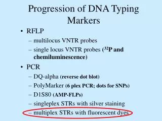
Progression of DNA Typing Markers. RFLP multilocus VNTR probes single locus VNTR probes ( 32 P and chemiluminescence) PCR DQ-alpha (reverse dot blot) PolyMarker (6 plex PCR; dots for SNPs) D1S80 (AMP-FLPs) singleplex STRs with silver staining multiplex STRs with fluorescent dyes.

E N D
Progression of DNA Typing Markers • RFLP • multilocus VNTR probes • single locus VNTR probes (32P and chemiluminescence) • PCR • DQ-alpha (reverse dot blot) • PolyMarker (6 plex PCR; dots for SNPs) • D1S80 (AMP-FLPs) • singleplex STRs with silver staining • multiplex STRs with fluorescent dyes
Changing Technologies Paradigm Shift: Restriction Fragment-Length Polymorphisms to Short Tandem Repeats • Five RFLP probes provide almost exclusive identity (~ 1 in 109 individuals) • RFLP requires a minimum of 25 ng of relatively undegraded DNA (1000 - 20,000 basepairs) • Short Tandem Repeats (STRs) only require ~ 1 ng DNA that can be partially degraded • Discrimination power: 5 RFLP probes equals ~ 12 STR loci
Which Suspect, A or B, cannot be excluded from potential perpetrators of this assault?
Fill with Polymer Solution Argon Ion Laser - + Inlet (cathode) Outlet (anode) 5-20 kV Capillary Electrophoresis (CE) 50-100 m x 27 cm Burn capillary window DNA Separation occurs in minutes... Data Acquisition and Analysis
Automated sample injection Automated gel pouring ABI PRISM® 310 Genetic Analyzer Capillary electrophoresis with multi-color detection capabilities
ABI Prism 310 Genetic Analyzer capillary Syringe with polymer solution Injection electrode Autosampler tray Outlet buffer Inlet buffer
Close-up of ABI Prism 310 Sample Loading Area Electrode Capillary Sample Vials Autosampler Tray
ABI 310 Result 9.3 allele: 1071 sec 10 allele: 1073 sec Mass Spec Result 9.3 allele: 203.3 sec 10 allele: 204.8 sec Allele 10 COfilerTM size: 187 bp MS size: 83 bp
Automated PCR Setup and Mass Spec Sample Preparation MWG Biotech RoboAmp 4200
Mass Spec Sample Plates PerSeptive Biosystems (100 positions) Bruker (384 positions)
Gels Time-of-Flight Mass Spectrometry Detector DNA separations occur in microseconds! DNA Reaction Products (Size separated and drifting to the detector) Drift Region Electric-Field Free Pulsed Laser Beam High-Density Sample Array Ion Extractor Acceleration Region X-Y sample control
Bruker BIFLEX III Time-of-Flight Mass Spectrometer Capable of fully automated data acquisition on 384 or more samples per plate