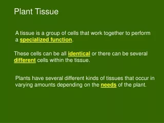
UNIT 4: PLANT TISSUE
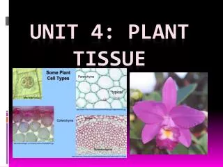
UNIT 4: PLANT TISSUE. MERISTEMATIC TISSUE. A flowering plant has the ability to grow its entire life because it possesses meristematic (embryonic) tissue.

UNIT 4: PLANT TISSUE
E N D
Presentation Transcript
MERISTEMATIC TISSUE • A flowering plant has the ability to grow its entire life because it possesses meristematic (embryonic) tissue. • The apical meristemare located at or near the tips of stems and roots, where they increase the length of their structures by means of mitosis. • This increase in length is called primary growth. • Monocots also have an intercalary meristem, this allows them to regrow lost parts. (It is found between mature tissues). EXAMPLE: GRASS CUT - GROW
Organization of a plant body MERISTEMATIC TISSUE AT TIP OF STEM MERISTEMATIC TISSUE AT TIP OF ROOTS
APICAL MERISTEM • Produces three types of meristems, and these develop into the three types of specialized primary tissues in the body of the plant: SPECIALIZED PRIMARY TISSUES: • Protoderm gives rise to the epidermis • Ground meristem produced ground tissue • Procambium produces vascular tissue
FUNCTIONS OF SPECIALIZED TISSUES • Epidermal tissue: forms the outer protective covering of a plant. • Ground tissue fills the interior of a plant. • Vascular tissue transports water and nutrients in a plant and provides support.
EPIDERMAL TISSUE • Single layer of closely packed, flat, brick shaped cells, with a large vacuole. • The aerial parts of the plant are covered with a cuticle. • Functions: - Cuticle minimizes water loss (because it has cutin) - Protects the plant against bacteria. Longitudinal section Cross section
SPECIALIZE EPIDERMAL CELLS TRICHOMES • ROOT HAIR • TRICHOMES • STOMA OF LEAF • CORK OF OLDER STEMS ROOT HAIRS STOMATA CORK CELLS
ROOT HAIRS • They are specialized epidermal cells of roots. • Unicellular outgrowth of the epidermal cell. • Functions: • Increase the surface area of the root for absorption of water and minerals. • Anchor the plant.
TRICHOMES CUTICLE • Specialized epidermal cell of stems and leaves. • Multicellularoutgrowths of the epidermis of stems and leaves. • Functions: • Protect the plant from sun • Conserve moisture. • Protect plant from herbivores, produce toxic substance. MULTICELLULAR
STOMATA • Specialized epidermal cells called guard cells, which are bean shaped, enclose an opening called the stoma or pore. • The guard cells contain a nucleus and chloroplasts. • It has a thick inner membrane and a thin outer membrane. • Woody plants have lenticels. • FUNCTIONS: • Transpiration • Gaseous exchange take place through the stomata. Thick inner membrane Thin outer membrane
CLOSED- NIGHT OPEN - DAY
PARENCHYMA COLLENCHYMA SCLERENCHYMA GROUND TISSUE
PARENCHYMA TISSUE • Occur in roots, stems and leaves. • Spherical, loosely packed, big, thin-walled cells with large vacuoles. • Intercellular airspaces between cells. FUNCTIONS: • If they have chloroplasts – photosynthesis. • If they have leucoplasts – they store products of photosynthesis. • They can divide to form more specialized cells
INTERCELLULAR AIR SPACES PARENCHYMA CELLS
COLLENCHYMA TISSUE • It is composed of unevenly thickened primary walls with additional cellulose and pectin deposits especially in the corners. • Found just beneath the epidermis of young stems. • The cells are slightly elongated, tightly packed and overlap each other. • FUNCTIONS: • Mechanical strengthening and support to plant organs
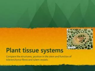
SCLERENCHYMA TISSUE • Cell walls have been thickened by impregnation with lignin. • The cell wall is evenly thickened and forms a waterproof barrier impermeable to water. • In the cell walls are pit canals that serve as channels between cells and to the outside world. • The lumen is small. • Two types of Sclerenchyma: • Stone cells and fibers FUNCTIONS: • Strengthening, support and protection. • Found in shell of nuts and hard parts of fruits • Fibers give rigidity and flexibility to the plant.
XYLEM PHLOEM Vascular tissue
XYLEM • Contains 2 types of conducting cells: tracheidsand vessel elements (VE). • Both cells are hollow and non-living but the VE is larger and has perforated plates in their end walls and are arranged to form a continuous vessel. • Tracheids have tapered ends with pits. • It also has other tissue: Xylem fibers, and Xylem parenchyma. • FUNCTION: Transports water and minerals from the roots to the leaves. Support and strengthening.
PHLOEM • Consist of sieve-tubes and companion cells. • The sieve-tubes form a continuous tube, they have cytoplasm but no nuclei. • They have sieve plates between cells. • The companion cell has a nucleus that controls both cells because they are connected by plasmodesmata. • It also has other tissue: Phloem fibers and phloem parenchyma • FUNCTIONS: Transports sucrose and other organic substances, including hormones, from the leaves to the roots. Support and strengthening.
METHODS TO STUDY CELLS • The microscope is an instrument designed to observe objects too small to be seen with the naked eye. • The human eye cannot distinguish objects much smaller than 0.1mm. • The microscope act as an extension of the eye, allowing one to see smaller objects.
ELECTRON MICROSCOPE • The electron microscope was developed in the late 1940’s and soon used in the study of cells. • Much more detail can be seen under an electron microscope. It can magnify parts of cells at least 300 000 times.
SCANNING ELECTRON MICROGRAPH TRANSMISSION ELECTRON MICROGRAPH