HUMAN DEFENSE MECHANISMS
HUMAN DEFENSE MECHANISMS. Categories of Defense Mechanisms. Physical barriers Skin and mucous membranes Chemical factors Mechanical factors Microbiological factors Innate immunity Adaptive immunity. Physical Barriers of Defense - Skin . Stratified squamous epithelium

HUMAN DEFENSE MECHANISMS
E N D
Presentation Transcript
Categories of Defense Mechanisms • Physical barriers • Skin and mucous membranes • Chemical factors • Mechanical factors • Microbiological factors • Innate immunity • Adaptive immunity
Physical Barriers of Defense - Skin • Stratified squamous epithelium • Chemical factors • Sebum (fatty secretion from sebaceous glands) • Lysozymes (perspiration produced by sweat glands) • Mechanical factors • Desquamation • Perspiration • Microbiological factors • Normal flora
Physical Barriers of Defense – Mucous Membranes • Columnar to squamous epithelium • Chemical factors • Lysozyme in tears, saliva and nasal secretions • Enzymes and HCl in stomach secretions • Defensins in small intestine • Mechanical factors • Lacrimal apparatus • Mucociliary clearance mechanism • Microbiological factors • Normal flora
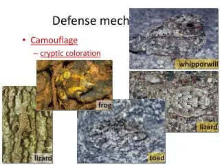
Normal Flora of Skin and Mucous Membranes • Population of microorganisms that may at any time be found residing on skin and mucous membranes of human host in the absence of disease • Skin • Staphylococcus epidermidis • Propionibacterium acnes • Corynebacterium species
Normal Flora of Mucous Membranes • Nasal mucosa • Staphylococcus aureus • Methicillin-susceptible (MSSA) • Methicillin-resistant (MRSA) • Nasopharyngeal mucosa • Streptococcus pneumoniae, Haemophilus influenzae, Moraxella catarrhalis • Buccal mucosa • Viridans streptococci, Neisseria species, Haemophilus species, Lactobacillus species, Prevotella species, Porphyromonas species, Fusobacterium species, Peptostreptococcus species
Normal Flora of Mucous Membranes • Colon mucosa • Bacteroides fragilis group, Clostridium species, Escherichia coli and other Enterobacteriaceae, Enterococccus species, Lactobacillus species, Candida albicans • Vaginal mucosa • Lactobacillus species, Gardnerella vaginalis, Mobiluncus species, Prevotella species, Porphyromonas species
PROBIOTICS • Definition • Food and Agriculture Organization of UN (FAO) and WHO • ‘live microorganisms which when administered in adequate amounts confer a health benefit on the host’ • Microorganisms • Bifidobacterium species • Lactobacillus bulgaricus • Lactobacillus casei • Streptococcus thermophilus
The Innate Response to Bacterial Pathogens Complement activation via alternative pathway Phagocytosis of pathogens by Macrophages Long-lived cells Secrete cytokines in innate and adaptive immunity Function as professional APC’s Neutrophils Historically called “microphages” Enter infected tissues in high numbers Short-lived cells
Activation of Tissue Macrophages • Activated macrophages initiate inflammatory response by secreting • Cytokines • Inflammatory mediators • Cytokines (chemoattractant cytokines / chemokines) • IL-1, IL-6, IL-8, IL-12 and TNF-alpha • Inflammatory mediators • Prostaglandins, leukotrienes, plasminogen activator, platelet-activating factor (PAF)
The Innate Response to Viral Pathogens • Virus infection of healthy cells results in production of • Interferon-alpha (IFN-alpha) • Interferon-beta (IFN-beta) • IFN-alpha and IFN-beta are type 1 interferons • Type 1 interferons • Inhibit virus replication • Activate natural killer (NK) cells • Increases expression of MHC-1 molecules
Natural Killer (NK) Cells • Large granular lymphocytes that circulate in blood • Functions • Killing infected cells (cytotoxic) • Secretion of cytokines • Activation by • Type 1 interferons • Infected cells • Stimulates cytotoxic function • IL-12 and TNF-alpha • Macrophages • Stimulates cytokine secretion
Natural Killer Cells • Activated NK cells release IFN-gamma which activates • Macrophages • Release IL-12 • Positive feedback system for NK and macrophages • Differentiate infected from uninfected cells • NK cells express receptors for MHC class I molecules • Binding of NK cells to MHC class I molecules turn off NK cells • NK cells provide innate immunity to intracellular pathogens
Adaptive Immune Response • Environment for starting provided by innate immune response • Consists of • Primary immune response • Follows initial exposure to antigen • Naive B and T cells • Establishment of memory • Secondary immune response • Follows second exposure to antigen • Memory B and T cells • Utilization of memory
Primary Immune Response • Begins with T cell activation and differentiation in secondary lymphoid tissue • CD4 TH1, CD4 TH2 and CD8 • Directed by cytokines • IL-12 and IFN-gamma (TH1) • IL-4 and IL-6 (TH2) • Continues with B cell activation in secondary lymphoid tissue • Cognate interaction with CD4 TH2 specific for same Ag
Role of T Cells in Primary Immune Response • Effector TH1 cells • Leave 2nd lymphoid tissue for infected tissue • Activate destruction of extracellular pathogens by macrophages • Effector CD8 cells • Leave 2nd lymphoid tissue for infected tissue • Kill infected cells • Effector TH2 cells • Remain in 2nd lymphoid tissue • Stimulates B cell differentiation into plasma cells
Role of B Cells in Primary Immune Response • Differentiation into plasma cells and antibody production • Locations for differentiation following CD4 TH2 cognate interaction • Medullary chords of lymph nodes • First wave of antibody secretion • Primary lymphoid follicles • Formation of germinal centers then migration to • Medullary chords of lymph nodes • Bone marrow • Second wave of antibody secretion
Secondary Immune Response • Adaptive immune response following second antigen exposure • Response is stronger and more rapid than primary • Classification • Short term (False) • 4 months or less following primary infection • Antibodies and effector T cells from naive lymphocytes • Long term (True) • 4 months or more following primary infection • Antibody and effector T cells from memory lymphocytes
Secondary Immune Response • No activation of naive B and T lymphocytes with specificity for pathogen • Mechanism for naive B cells • Suppression by • Immune complex (IC) of pathogen and IgG • IC’s bind to naive B cell • Receptor • Inhibitory Fc receptor (Fc-gammaRIIB1)
Clinical Application of Memory B Cell Activation • Prevention of • Hemolytic disease (anemia) of newborn • Hemolytic disease of newborn • Rh- mother with Rh+ fetus • Fetal RBC enter maternal circulation • No intervention • Maternal antibody against fetal RBC • Intervention with Anti-Rh, IgG (Rhogam) • No maternal antibody against fetal RBC
Immunological Memory and a Variant Pathogen • Infection with Influenza viruses • Influenza A and B viruses mutate surface antigens • Antigenic drift (A and B) • Antigenic shift (A) • Viral strategy • Erosion of protective immunity • Strategy of immune system • Respond to strains with epitopes previously encountered • IM limited to epitopes shared by infecting and original strain