Download
1 / 40
410 likes | 686 Vues
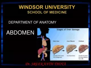
DEPARTMENT OF ANATOMY. WINDSOR UNIVERSITY SCHOOL OF MEDICINE. ABDOMEN. Dr. SREEKANTH THOTA. Liver. The liver is the largest gland in the body and, after the skin, the largest single organ. It weighs approximately 1500 g.

E N D
DEPARTMENT OF ANATOMY WINDSOR UNIVERSITYSCHOOL OF MEDICINE ABDOMEN Dr. SREEKANTH THOTA
Liver The liver is the largest gland in the body and, after the skin, the largest single organ It weighs approximately 1500 g It occupies almost all of the right hypochondrium, epigastrium and left hypochondrium
Surfaces, Peritoneal Reflections, and Relationships of the Liver
Surfaces • The liver has a convex diaphragmatic surface (anterior, superior, and some posterior) and a relatively flat or even concave visceral surface (posteroinferior), which are separated anteriorly by its sharp inferior border.
Peritoneal Reflections • Diaphragmatic surface • 1. Falciform ligament • 2. right triangular ligament • 3. left triangular ligament • 4. coronary ligament The diaphragmatic surface of the liver is covered with visceral peritoneum, except posteriorly in the bare area of the liver where it lies in direct contact with the diaphragm.
Peritoneal Reflections • Visceral surface of the liver • 1. ligamentum teres or round ligament • 2. ligamentum venosum: fibrous remnant of the fetal ductus venosus, which shunted blood from the umbilical vein to the IVC, short-circuiting the liver.
Anatomical Lobes of the Liver • The liver is divided into right and left lobes by fossae for the gallbladder and the inferior vena cava. • The quadrate and caudate lobes are described as arising from the right lobe of liver, but functionally are distinct.
Porta hepatis • Opening in the Rt lobe • Between the caudate and quadrate lobes • Transmits the following • 1. portal vein(Rt.& Lt. Branches) • 2. hepatic duct (Rt.& Lt.branches) • 3. hepatic artery (right and left branches) • 4. Lymphatics & nerves
Blood Vessels of the Liver • The liver, like the lungs, has a dual blood supply (afferent vessels): a dominant venous source and a lesser arterial one. • The portal vein brings 75 to 80% of the blood to the liver. • Arterial blood from the hepatic artery, accounting for only 20 to 25%.
Hepatic veins • The hepatic veins, formed by the union of collecting veins that in turn drain the central veins of the hepatic parenchyma open into the IVC just inferior to the diaphragm. • Are usually 3 in number(right, intermediate (middle), and left hepatic veins)
Biliary Ducts • The biliary ducts convey bile from the liver to the duodenum. • The right and left hepatic ducts drain the right and left (parts of the) liver, respectively. • Shortly after leaving the porta hepatis, the right and left hepatic ducts unite to form the common hepatic duct, which is joined on the right side by the cystic duct to form the bile duct.
Bile Duct • The length of the bile duct varies from 5 to 15 cm, depending on where the cystic duct joins the common hepatic duct. • In the first part:it lies in front of the right margin of the portal vein and on the right of the hepatic artery. • In the second part of its course, it is situated behind the first part of the duodenum to the right of the gastroduodenal artery.
In the third part of its course, it lies in a groove on the posterior surface of the head of the pancreas . Here, the bile duct comes into contact with the main pancreatic duct. • The bile duct ends below by piercing the medial wall of the second part of the duodenum about halfway down its length.
It is usually joined by the main pancreatic duct, and together they open into a small ampulla in the duodenal wall, called the hepatopancreatic ampulla (ampulla of Vater). • The ampulla opens into the lumen of the duodenum by means of a small papilla, the major duodenal papilla
Gallbladder • The gallbladder is a pear-shaped sac lying on the undersurface of the liver . • 7 to 10 cm long. • The pear-shaped gallbladder can hold up to 50 mL of bile.
The gallbladder has three parts • 1. Fundus • 2. Body: • 3. Neck:
Blood Supply • The cystic artery, a branch of the right hepatic artery supplies the gallbladder. • The cystic vein drains directly into the portal vein.
Gallstones • A gallstone is a concretion in the gallbladder, cystic duct, or bile duct composed chiefly of cholesterol crystals. • The distal end of the hepatopancreatic ampulla is the narrowest part of the biliary passages and is the common site for impaction of gallstones. • The infundibulum of the gallbladder is another common site for impaction.
Gallstones in the Duodenum • Because of their proximity to the gallbladder, the superior part of the duodenum and the transverse colon are most likely to develop a fistula. • A large gallstone entering the small intestine in this way may become trapped at the ileocecal valve, producing a bowel obstruction (gallstone ileus).
Acute Cholecystitis Acute cholecystitis is a sudden inflammation of the gallbladder that causes severeabdominal pain.
Acute cholecystitis produces discomfort in the right upper quadrant or epigastrium. Inflammation of the gallbladder may cause irritation of the subdiaphragmatic parietal peritoneum, which is supplied in part by the phrenic nerve (C3, 4, and 5). This may give rise to referred pain over the shoulder, because the skin in this area is supplied by the supraclavicular nerves (C3 and 4). Boas' or Boas's sign is hyperaesthesia below the right scapula can be a symptom in acutecholecystitis (inflammation of the gallbladder)
Murphy's sign • Typically, it is positive in cholecystitis. • It is performed by asking the patient to breathe out and then gently placing the hand below the costal margin on the right side at the mid-clavicular line (the approximate location of the gallbladder)
Portal Vein and Portal Systemic Anastomoses • Portal systemic anastomoses, in which the portal venous system communicates with the systemic venous system.
Portosystemic Shunts • A common method for reducing portal hypertension is to divert blood from the portal venous system to the systemic venous system by creating a communication between the portal vein and the IVC. • Join the splenic vein to the left renal vein, after splenectomy (splenorenal anastomosis or shunt)