Gastroenterology Chapter 3
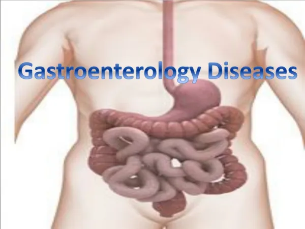
Gastroenterology Chapter 3. Anatomy of the GI System:. - Upper gastrointestinal system includes: Oral Cavity & Pharynx Esophagus Stomach -Lower gastrointestinal system includes: Small Intestines large Intestines. Physiology of the GI System:. Upper gastrointestinal system:

Gastroenterology Chapter 3
E N D
Presentation Transcript
Gastroenterology Chapter 3
Anatomy of the GI System: -Upper gastrointestinal system includes: Oral Cavity & Pharynx Esophagus Stomach -Lower gastrointestinal system includes: Small Intestines large Intestines
Physiology of the GI System: Upper gastrointestinal system: 1. Begins the digestion of nutrients. Lower gastrointestinal system: 2. Finish the digestion of nutrients 3. Nutrient absorption 4. Prepare non-digestible food for waste removal
Anatomy Specifics: Oral Cavity Physical (Mechanical) Digestion: -Mouth: Contains the teeth; tongue; hard palate; and soft palate, & uvula. -The teeth tear, chew, and grind the food during the process of mastication. -The tongue moves food toward the teeth and mixes food with saliva. Chemical Digestion: -Saliva (contains enzymes) secreted by the three salivary glands: parotid glands, sublingual glands submandibular glands -The main enzyme (salivary amylase) chemically begins the digestion of carbohydrates.
Pharynx: -Swallowing or deglutition moves food into the throat or pharynx. -When food is swallowed, the epiglottis closes the entrance to the larynx, so that food doesn’t pass into the trachea.
Esophagus: -A flexible, muscular tube that connects the pharynx to the stomach. -food is able to pass through the esophagus by muscular contractions called peristalsis.
Stomach: -Divided into four areas: 1. cardia 2. fundus 3. body 4. pylorus -Function: digest proteins. -The gastric mucosa (interior lining) is arranged in thick, deep folds known as rugae which expand as the stomach fills with food. -Sphincter Muscles: lower esophageal and pyloric
Small Intestine: -The small intestine is a long, hollow tube that receives chyme from the stomach. -around 22-25 feet long -It is divided into three regions: 1. duodenum 2. jejunum 3. ileum -lined with villi to increase surface area for nutrient absorption; villi secrete lactase for digestion of carbs. Functions: finish nutrient digestion; nutrient absorption.
Large Intestine: -A larger diameter hollow tube that receives undigested material and water from the small intestine. -about 6-7 feet long -Regions: 1. cecum 2. appendix 3. colon* 4. rectum 5. anus -Four quadrants of the colon: ascending, transverse, descending , and sigmoid. Functions: prepare undigested food for removal from the body; reabsorb as much water as possible; bacteria synthesize vitamin K.
Accessory Organs: part of the digestive system, but food does not travel to these organs. Liver: -The liver is the largest solid organ in the body, located in the upper right abdominal cavity. -Function: produces bile; stores glycogen; acts as a filter for your blood; regulates glucose and amino acids; makes plasma proteins & blood clotting factors. Gallbladder: -Is found posterior to the liver; sits underneath it. -Function: concentrates and stores bile from the liver; sends bile to the small intestine through the common bile duct.
Pancreas: -Is found posterior to the stomach. -Function: secrete digestive enzymes into the pancreatic duct which leads to the duodenum;also is an organ of the endocrine system.
Physiology of Digestion There are two parts to digestion: 1. Mechanical 2. Chemical (enzymes) -Mechanical Digestion: mastication, deglutition, and peristalsis to break down foods.
Chemical Digestion - Enzyme Action: Oral Cavity: Salivary Amylasein saliva begins the digestion of carbohydrates. Stomach: digestion of proteins. 1. Gastrin- hormone causes the release of HCl & Pepsinogen. 2. Hydrochloric Acid (HCl) mixes with Pepsinogen to make Pepsin.
Small Intestine: all chemical digestion is completed. 1. Cholecystokinin (hormone) stimulates the gallbladder to release bile which is sent to the small intestines for lipid (fat) digestion. 2. Pancreas secretes enzymes and sends them to the duodenum: Amylase- carbs Lipase- lipids Protease- proteins Nuclease- nucleic acids
Abdomen and AbdominopelvicCavityStructures: -The walls of the abdominopelvic cavity are lined by peritoneum, a membrane that secretes peritoneal fluid. -Omentum is part of the peritoneum which supports the stomach and covers the small intestine. -The peritoneal fluid fills spaces between the organ; allows them to slide past each other. -The blood supply to the stomach, small intestine, liver, gallbladder, and pancreas comes from the celiac trunkof the aorta.
Alimentary Canal & Accessory Structures Figure 3-7 Gastrointestinal system. (Robert W. Ginn/PhotoEdit Inc.) End of Test #1 Material.
Ch. 3 Gastroenterology:Diseases and Conditions Eating -Anorexia -Dysphagia -Polyphagia Mouth and Lips -Cheilitis -Sialolithiasis -Stomatitis -Glossitis
Figure 3-8 Glossitis (Centers for Disease Control and Prevention [CDC])
Esophagus and Stomach -Dyspepsia -Esophageal varices -Gastritis -Gastroenteritis -Gastroesophageal reflux disease (GERD) -Heartburn -Hematemesis -Nausea and vomiting (N&V) -Peptic ulcer disease (PUD) -Stomach cancer
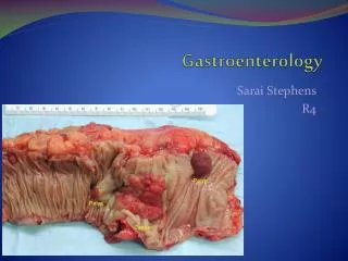
Figure 3-10 Gastric ulcer (David M. Martin, M.D./Photo Researchers, Inc.)
Duodenum, Jejunum, Ileum -Ileus -Intussusception -Volvulus
Cecum and Colon -Appendicitis -Colic -Colon cancer -Diverticulum -Dysentery -Gluten enteropathy -Inflammatory bowel disease (IBD) -Irritable bowel syndrome (IBS) -Polyp
Figure 3-12 Diverticula (David M. Martin, M.D./Photo Researchers, Inc.)
Figure 3-15 Colonic polyps (Staats/Custom Medical Stock Photo, Inc.)
Rectum and Anus -Hemorrhoids -Proctitis -Rectocele
Defecation and Feces -Constipation -Diarrhea -Flatulence -Hematochezia -Incontinence -Steatorrhea
Abdominal Wall and Abdominal Cavity -Adhesions -Hernia -Peritonitis
Figure 3-17 Peritonitis (Custom Medical Stock Photo, Inc.)
Liver -Ascites -Cirrhosis -Hepatitis -Hepatomegaly -Jaundice -Liver Cancer
Liver Disease: Hepatitis is the most common chronic liver disease. -Hepatitis A -Hepatitis B -Hepatitis C -Hepatitis D -Hepatitis E
Figure 3-20 Jaundice (Dr. M.A. Ansary/Photo Researchers, Inc.)
Figure 3-21 Liver cancer (Gca/Photo Researchers, Inc.)
Gallbladder and Bile Ducts -Cholangitis -Cholecystitis -Cholelithiasis
Pancreas -Pancreatic cancer -Pancreatitis
Laboratory and Diagnostic Procedures: Blood Tests -Albumin -Alkaline phosphatase (ALP) -ALT and AST -Bilirubin -GGT -Liver function tests (LFTs)
Gastric and Feces Specimen Tests -CLO test -Culture and sensitivity (C&S) -Fecal occult blood test -Gastric analysis -Ova and parasites (O&P) Radiologic Procedures -Barium enema -Cholangiography -Computerized axial tomography -Flat plate of the abdomen -Gallbladder ultrasound -Magnetic resonance imaging (MRI) scan -Oral cholecystography (OCG) -Upper gastrointestinal series (UGI)
Medical and Surgical Procedures: Medical Procedures -Insertion of nasogastric tube
Surgical Procedures -Abdominocentesis -Appendectomy -Biopsy -Bowel resection and anastomosis -Cholecystectomy -Choledocholithotomy -Colostomy
Figure 3-28 Colostomy and stoma (Pearson Education/PH College)
Surgical Procedures -Endoscopy -Exploratory laparotomy -Gastrectomy -Gastroplasty -Gastrostomy -Hemorrhoidectomy -Herniorrhaphy -Jejunostomy -Liver transplantation -Polypectomy
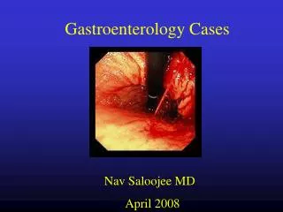
Endoscopic Procedures -Esophagoscopy -Gastroscopy -Esophagogastroduodenoscopy (EGD) -Sigmoidoscopy -Colonoscopy