Download
1 / 29
290 likes | 312 Vues
Axiva Sichem Biotech specializes in manufacturing quality lab filtration products and associated products for laboratory applications. Our company has continued to supply and introduce innovative products to the market in many countries. We offer the widest range of filter papers, syringe filters, membrane filters, pipettes, glass fiber filters, filtration assembly, filter funnel, biosafety cabinets, laminar flow cabinets and many other laboratory equipmentu2019s. http://www.axivasichem.com/blotting-membranes.aspx

E N D
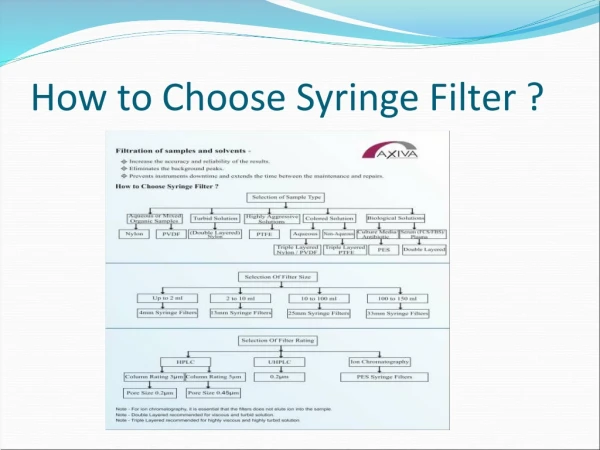
SYRINGE FILTERS A syringe filters generally consists of a plastic housing with a membrane which serves as a filter. The fluid to be purified may be cleaned by drawing it up the syringe through the filter, or by focusing the unfiltered fluid out through the filter. Membrane Filters Membrane Filters or membrane is microporus films with specific filtration rating. Membrane retains particles and micro organisms that exceed their filtration rating by acting as a physical barrier and capturing such particles on the surface of membrane.
Case study Chloroform (Halogenated Hydro carbon) DMSO (Dimethyl Sulfoxide) Lipid solution Hormones IVF / NMR Non – aqueous colored solutions Highly viscous aqueous solution (Plasdone XL, kolldon SR, Xanthan gum, Avicel PH 112)
What is blotting ? Blotting are techniques for transferring DNA, RNA and proteins onto a carrier so they can be separated, and often follows the use of a gel electrophoresis. The southern blot is used for transferring DNA, the Northern blot for RNA and the western blot for PROTEIN.
TYPES OF BLOTTING TECHNIQUES Blotting Technique Southern Blot Western Blot Northern Blot It is used to detect DNA. It is used to detect protein. It is used to detect RNA.
Protein Blotting Protein blotting is an analytical Method that involves the immobilization Of proteins on membranes before detection Using monoclonal or polyclonal antibodies. There are different blotting protocols (dot blot, 2D blot); one of the most powerful is western blotting.
Principle of Western Blotting Western blotting is an Immunoblotting technique which rely on the specificity of binding between a molecule of interest and a probe to allow detection of the molecule of interest in a mixture of many other similar molecules. In Western blotting, the molecule of interest is a protein and the probe is typically an antibody raised against that particular protein. The SDS PAGE technique is a prerequisite for Western blotting.
Flow Diagram of Western Blot Protein Blot on SDS Polyacrylamide Nitrocellulose Gel Electrophoresis
Label With Specific Detect Antibody Antibody Reveals Protein of Interest
Western Blot (Immunoblotting) A technique for detecting specific proteins separated by electrophoresis by use of labeled antibodies. So called Since it has some similarity to a southern blot.
Definition The Western Blot is an analytical technique used to detect specific proteins in a given sample of tissue homogenate or extract.
Advantages of Western Blot Western blot analysis can analyze any protein sample whether from cells or tissues, but also can analyze recombinant proteins synthesized in vitro. Western blot is dependent on the quality of antibody you use to probe for your protein of interest, and how specific it is for this protein
Gel Electrophoresis Uses gel electrophoresis to separate native or denatured proteins by the length of the polypeptide (denaturing conditions) or by the 3-D structure of the protein (native / non-denaturing conditions). The proteins are then transferred to a membrane (typically nitrocellulose or PVDF), where they are probed (detected) using antibodies specific to the target protein.
Monoclonal and Polyclonal Antibodies are used Here are now many reagent companies that specialize in providing antibodies (both monoclonal and polyclonal antibodies ) Against tens of thousands of different proteins.
Most Uselful In… This methods is used in the fields of molecular biology, Biochemistry, immunogenetics and other molecular biology disciplines.
The Procedure Includes Tissue Preparation Gel Electrophoresis Transfer Blocking Detection Analysis
Samples may be taken from whole tissue or from cell culture. In most cases, solid tissues are first broken down mechanically using a blender. It should be noted that bacteria, virus or environmental samples can be the source of protein and thus Western blotting is not restricted to cellular studies only. Assorted detergents, Salts, and buffers may be employed to encourage lysis of cells and to solubilize proteins. Tissue preparation is often done at cold temperature to avoid protein denaturing.
Gel Electrophoresis The proteins of the samples are separated using gel electrophoresis. Separation of proteins may be by isoelectric point molecular weight, electric charge, or a combination of these factors. The Principle involved is the difference in the ELECTROPHORETIC MOBILITIES of different proteins.
Transferring In Order to make the proteins accessible to antibody detection, they are moved from within the gel onto a membrane made of nitrocellulose or polyvinylidene difluoride (PVDF). The membrane is placed on top of the gel, and a stack of filter papers placed on top of that. The entire stack is placed in a buffer solution which moves up the paper by capillary action, bringing the proteins with it. Another method for transferring the proteins is called electro blotting and uses an electric current to pull proteins from the gel onto the PVDF or nitrocellulose membrane.
Blocking The membrane has the ability to bind to proteins in this case both the target and antibodies are proteins and so there could be some unwanted binding. Blocking of non-specific binding is achieved by placing the membrane in a dilute solution of protein – typically Bovine serum albumin (BSA) with a minute percentage of detergent such as tween 20. The protein in the dilute solution attaches to the membrane in all places where the target proteins have not attached. Thus, when the antibody is added, there is no room on the membrane for it to attach other than on the binding sites of the specific target protein.
Detection During the detection process, the membrane is “Probed” for the protein of interest with a modified antibody which is linked to a reporter enzyme, which when exposed to an appropriate substrate drives a colorimetric reaction and produces a color.
Analysis After the unbound probes are washed away, the western blot is ready for detection of the probes, that are labeled and bound to the protein of interest. Size approximations are taken by comparing the stained bands to that of the marker loaded during electrophoresis. The process is repeated for a structural protein, such as actin, or tubulin that should not change between samples.
Advantages While ELISA being a non-specific test, Western blotting is a more specific test for detection of HIV. It can detect one protein in a mixture of proteins while giving information about the size of the protein and so is more specific. Western blot test is referred to as the ‘Gold standard’ It also tells you how much protein has accumulated in cells.
Western Blot in Clinical Medicine The confirmatory HIV test employs a Western blot to detect anti-HIV antibody in a human serum sample. Proteins from known HIV-infected cells are separated and blotted on a membrane then, the serum to be tested is applied in the primary antibody incubation step; free antibody is washed away, and a secondary anti-human antibody linked to an enzyme signal is added . The stained bands then indicate the proteins to which the patient’s serum contains antibody. A western blot is also used as the definitive test for Bovine Spongiform encephalopathy (BSE, commonly referred to as ‘mad cow disease’) Some forms of Lyme disease testing employ Western Blotting.
Western Blot a Confirmatory test in HIV Infection The virus is enveloped with different The detection of these proteins are useful in the detection of the presence of the virus. Western Blotting helps in the detection of these proteins.
Contact Us Manufacturing Unit - 424,EPIP, Industrial Estate, Sector-53, Phase-III, Kundli, Sonipat Haryana-131028 (INDIA) Marketing Office- RU-46, Pitampura, Delhi-110034 (INDIA) Phone No - +91-11-40578224 / 25 / 26 Mobile No - +91-8800493923 Website- www.axivasichem.com