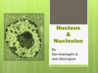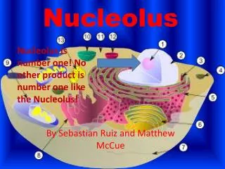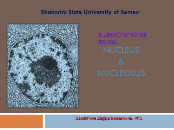

nucleolus
E N D
Presentation Transcript
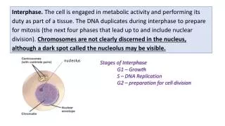
Interphase. The cell is engaged in metabolic activity and performing its duty as part of a tissue. The DNA duplicates during interphase to prepare for mitosis (the next four phases that lead up to and include nuclear division). Chromosomes are not clearly discerned in the nucleus, although a dark spot called the nucleolus may be visible. nucleolus Stages of Interphase G1 – Growth S – DNA Replication G2 – preparation for cell division
Prophase. (1)Chromatin in the nucleus begins to condense and becomes visible in the light microscope as chromosomes. (2)The nuclear membrane dissolves, marking the beginning of late prophase (prometaphase). (3) Proteins attach to the centromeres creating the kinetochores. Microtubules attach at the kinetochores and the chromosomes begin moving. (1) Chromatin in the nucleus begins to condense and becomes visible in the light microscope as chromosomes Centrosomes with centriole pairs Microtubules – beginning of the Spindle (2)The nuclear membrane dissolves, marking the beginning of prometaphase.
Metaphase. Spindle fibers align the chromosomes along the middle of the cell nucleus. This line is referred to as the metaphase plate. This organization helps to ensure that in the next phase, when the chromosomes are separated, each new nucleus will receive one copy of each chromosome. Spindle Chromosome Metaphase plate (mid-line)
Anaphase. The paired chromosomes separate at the kinetochores and move to opposite sides of the cell. Motion results from a combination of kinetochore movement along the spindle microtubules and through the physical interaction of polar microtubules. The paired chromosomes separate at the kinetochores and move to opposite sides of the cell. Spindle microtubules
Telophase. New membranes form around the daughter nuclei while the chromosomes disperse and are no longer visible under the light microscope. Cytokinesis or the partitioning of the cell may also begin during this stage. New membranes form around the daughter nuclei while the chromosomes disperse and are no longer visible under the light microscope Cytokinesis. In animal cells, cytokinesis results when a fiber ring composed of a protein called actin around the center of the cell contracts pinching the cell into two daughter cells, each with one nucleus. In plant cells, the rigid wall requires that a cell plate be synthesized between the two daughter cells