Introduction to glaucoma
Introduction to glaucoma. From a Clinician’s Standpoint. Readings. Alexander, pp. 258-276 Kanski Perimetry, pp. 24-32 OCT, HRT & GDx, pp. 47-52 Glaucoma, pp. 371-393. Overview. Introduction Diagnosis History and risk factors Tonometry Pachymetry Gonioscopy ONH structure

Introduction to glaucoma
E N D
Presentation Transcript
Introduction to glaucoma From a Clinician’s Standpoint
Readings • Alexander, pp. 258-276 • Kanski • Perimetry, pp. 24-32 • OCT, HRT & GDx, pp. 47-52 • Glaucoma, pp. 371-393
Overview • Introduction • Diagnosis • History and risk factors • Tonometry • Pachymetry • Gonioscopy • ONH structure • ONH function • Risk Assessment • Management • Adherence • Communication
Glaucoma introduction • Optometric lifelong learning • Optic nerve head and nerve fiber layer imaging • IOP and pachymetry • Pilocarpine Timoptic Xalatan
Diagnosis • History and risk factors • Asymptomatic until late • Age • Race: younger age at diagnosis, increased risk of blindness (African-Americans) • Hispanics especially over the age of 75 • Family history • ? Myopia, Diabetes, hypertension
Diagnosis • Tonometry (IOP) • Goldmann applanation • Single most important risk factor • For development of glaucomatous optic neuropathy • Risk increases with increasing IOP • Vital for initial diagnosis and management • Currently, only major treatable risk factor
Diagnosis • Tonometry (IOP) • Glaucoma may develop at any IOP level • Central corneal thickness data via pachymetry to predict the “true” IOP • More to the story than adjusting for central corneal thickness • Biomechanical influences on the cornea influence the IOP….. • New tonometers: Rechart Ocular Response Analyzer, Pascal tonometer, Icare
Diagnosis Pachymetry Measurement of corneal thickness Normal population mean value: 544 + 34 microns Corneal thickness is a risk factor for IOP independent of its ability to skew IOP Thinner than normal increases risk Can also make adjustment for IOP: most corrections based on values between 2.5-3.5 mm Hg per 50 microns of CCT (central corneal thickness) from 545 microns
Diagnosis • Gonioscopy • Careful exam of anterior chamber angle • My lens of choice • Essential for glaucoma suspects and diagnosis • POAG: Primary Open Angle Glaucoma • Permits exclusion of angle closure and other secondary types of glaucoma
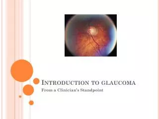
Diagnosis • ONH Structure • Evaluation of the ONH and retinal nerve fiber layer (RNFL) • View the ONH and RNFL • Also ONH and RNFL Imaging: OCT, HRT, GDx • Structural damage prior to VF defects • 50% rule • The BEST way: slit lamp retinal biomicroscopy through a dilated pupil allowing for magnified stereoscopic view • To view the ONH
DIAGNOSIS • ONH Structure • Evaluation of the neuroretinal rim (NRR) • Healthy rim tissue quadrants thickest thinnest • Inferior • Superior • Nasal • Temporal
DIAGNOSIS • ONH Structure • Focal thinning of the rim tissue or “notch” • Vertical cup elongation • Increased cup depth • Disc asymmetry • Disc hemorrhage • Cup size
DIAGNOSIS • RNFL • Red-free filter view • Better: RNFL Imaging • SLGT: Scanning Laser Glaucoma Test • Standard of Care? To Manage Glaucoma • HRT: Heidelberg Retina Tomograph • Scanning laser tomography for the optic nerve • GDx VCC • Scanning laser polarimetry for the RNFL • OCT: Optical Coherence Tomography • For the optic nerve and RNFL
DIAGNOSIS • Scanning Laser Glaucoma Tests • All have excellent diagnostic performance for early glaucoma • Not meant to be used in isolation
DIAGNOSIS • ONH Function • Measuring the Visual Field (VF) via standard automated perimetry • Humphrey Field Analyzer • Central threshold • 30-2 • 24-2 • SITA
DIAGNOSIS • ONH Function • VF Defects • Reflect damage to NFL bundles as they track toward the optic nerve • ONH appearance should match the VF • Progression of defects: paracentral / nasal step arcuate • Central vision not affected until late in course
DIAGNOSIS • ONH Function • Additional VF Instruments • FDT (Frequency Doubling Technology) Matrix • SWAP (Short-wavelength automated perimetry) or blue-yellow perimetry • Heidelberg Edge Perimeter
Risk assessment • Who to treat? • When to treat? • What extent to treat? • Currently review results as a whole • Detect any damage • Set target IOP to reduce or eliminate further damage • Monitor every 3 months
Risk Assessment • The STAR Risk calculator • Pfizer, Inc. • Age. IOP, CCT, vertical C/D ratio, pattern standard deviation (PSD) for a HFA II threshold visual field, diabetes status • Takes into account many important trials and studies related to glaucoma
Management • Classes of medical agents • Prostaglandin analogs (Xalatan, Lumigan, Travatan) • Beta-adrenergic antagonists (Timoptic, Betoptic-S) • Carbonic Anhydrase Inhibitors (Trusopt, Azopt) • Adrenergic Agonists (Alphagan-P) • Cholinergic Agonists (Pilocarpine) • What I like to ask in clinic?…..MOA’s, side effects, contraindications, how to write the Rx
MaNAGEMENT • Medical Agents…considerations • How well do they work? (% drop in IOP) • Chances of compliance? • Cost issues? • Prostaglandins have become the primary agents of therapy…others as adjuncts when needed
MANAGEMENT • Laser and surgery • SLT: Selective Laser trabeculoplasty • ALT: Argon Laser trabeculoplasty • Trabeculectomy
MANAGEMENT • Overview of Monitoring $ • Every 3 months • yearly DFE, VF, SLGT • Pachymetry at diagnosis • Photos and gonioscopy every 2 years
Adherence • Nonadherence to topical glaucoma meds • Prevalent • Difficult to detect • Patients must understand progression and be concerned about future consequences of glaucoma • Active learners and participants tend to adhere better
Communication…a few suggestions • 1. Tell me how you have been taking your medications? • 2. Tell patient you will not be judgemental regarding non-adherence • 3. Explain how information about adherence will affect treatment decisions • 4. Ask about forgetting or missing medications • 5. non-adherent…tell me what you understand about glaucoma and what your concerns are