Download
1 / 66
660 likes | 827 Vues
REGULATION OF ACIDS, BASES, & ELECTROLYTES. Key Topics. Ventilation as an Acid-Base Controller Renal Anatomy Review Starling’s Law of the Capillary Fluid and Electrolyte balance Renal Physiology Kidney as an Acid-Base Controller Diuretics. Ventilation Control & Regulation.

E N D
Key Topics • Ventilation as an Acid-Base Controller • Renal Anatomy Review • Starling’s Law of the Capillary • Fluid and Electrolyte balance • Renal Physiology • Kidney as an Acid-Base Controller • Diuretics
Ventilation Control & Regulation • Volatile acids (i.e. Carbonic Acid) are removed via the lungs. • The volume of gas that is removed is directly related to the alveolar ventilation. • The amount of alveolar ventilation is determined by the control and regulation of ventilation.
Ventilation Control • Primary respiratory generator is the inspiratory & expiratory centers in the medulla. • These are in turn regulated by: • The apneustic center • The pneumotaxic center • The cerebral cortex – voluntary changes in ventilation
Ventilation Regulation • Amount of ventilation is also regulated through feedback mechanisms. • Chemoreceptors • Central and Peripheral • Reflex Mechanisms • Hering-Breuer • J Receptors
Central Chemoreceptors • Located in the medulla of the CNS • Not to be confused with the respiratory centers! • Chemoreceptors are bathed in CSF that respond to the pH of the CSF ( H+, ventilation). • CSF is relatively impermeable to ions (i.e. HCO3-), only gases (i.e. PaCO2). • Known as the blood-brain barrier. • Slight changes in PaCO2 will yield a change in ventilation. • No response to hypoxemia.
Peripheral Chemoreceptors • Located in arterial system • Carotid Bodies • Aortic Bodies • They respond to the blood passing by them. • Both respond to: • PaCO2/pH: Large increases are needed in PaCO2 or pH before changes in minute ventilation occur. • PaO2: Comes into play with chronic pulmonary disease (hypoxic drive) • Dramatic increase in stimulation when PaO2 < 60 mm Hg. • Baroreceptors: Regulate blood pressure & 2° Ventilation.
Regulation of Ventilation and Blood Gases in Progressive Pulmonary Disease
Reflexes • Hering-Breuer • Stretch reflex • Regulates tidal volume & respiratory rate to minimize work of breathing. • Active during overinflation and underinflation. • Inflation reflex • Deflation reflex (rapid-shallow breathing) • J receptors • Juxtapulmonary capillary receptors • Interstitial tissue of the A-C membrane • Respond to thickening of this membrane (? pressure) • Pulmonary Edema & Pulmonary Fibrosis • Result in tachypnea & hyperventilation
Renal Function • Three primary functions • Excretion of nonvolatile waste products (including nonvolatile acids) • Regulation of blood volume. • Regulation of various electrolytes & blood constituents.
Kidney A & P • Retroperitoneal organ. • Outer cortex; inner medulla. • Ureter transports urine from kidney to bladder.
Nephron • Functional unit of the kidney. • 1,300,000 per kidney • Composed of • Bowman’s Capsule • Proximal Convoluted Tubules • Loop of Henle • Distal Convoluted Tubules • Collecting Tubule • EGAN
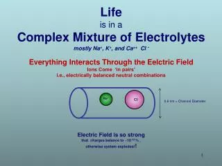
Fluid Compartments • Total amount of water in 70 kg man = 40 L • Accounts for 60% of the body’s weight • Intracellular (within the cells): 25 L • Red Blood Cell volume: 2 L • Extracellular (outside the cells): 15 L • Interstitial (in the spaces between cells): 12 L • Plasma: 3 L • 2/3rd of the blood volume is in the cell.
Electrolytes • Two types of substances found in body water: • Non-Electrolytes: Intact uncharged particles • Urea, Creatinine, Glucose • Electrolytes: Particles that dissociate and carry electrical charges • Cations – Positively charged • Anions – Negatively charged
Capillary Structure & Function • Capillaries allow movement of substances (O2, K+, Glucose) out of the capillary and into the cell through diffusion and movement of water by osmosis. • Capillaries have (basement) membranes with varying permeability. • Different organs allow substances of various size to diffuse across. (lung vs. kidney vs. intestine) • Some substances never leave the vascular space under normal conditions (hemoglobin, albumin)
Capillary Structure & Function • Capillaries are like a leaky hose; the bulk of the fluid continues along the hose but some fluid leaks out due to pressure within the hose.
Fluid Dynamics in the Capillary • Five factors regulate fluid movement in and out of the capillary. • 2 Hydrostatic pressures • One inside & One outside • 2 Osmotic (Oncotic) pressures • One inside & One outside • The relative permeability of the capillary wall
Hydrostatic Pressure • HYDROSTATIC PRESSURES PUSH FLUID OUT. • Increased capillary (arterial side) hydrostatic pressure forces fluids out of the capillary. • Increased interstitial hydrostatic pressure forces fluid out of the interstitial space (into the alveolus in fulminate pulmonary edema)
Osmotic Pressure • OSMOTIC PRESSURES PULL FLUID IN. • Plasma proteins (hemoglobin, albumin) [aka colloids] that don’t move out of the vascular space and cross the capillary wall exert an osmotic (oncotic) pressure within the vessel and draw fluid (and any associated soluble waste products) back into the capillary at the venous end. • 2 Osmotic pressures (capillary and interstitial)
Starling Forces • 5th factor is the permeability of the membrane. • This phenomenon is known as Starling forces. • This movement of fluid allows for: • Delivery of nutrients to cell • Waste products to be reabsorbed • Fluid and Electrolyte Homeostasis both in peripheral cells and in the kidney.
Net Effect • Excess fluid left in the tissues can be caused by: • Increased Capillary Hydrostatic pressure • Decreased Capillary Osmotic pressure • Decreased Interstitial Hydrostatic pressure • Increased Interstitial Osmotic pressure • Increased Permeability of the capillary wall
Bowman’s Capsule • Located in cortical portion of kidney. • Beginning of the Proximal Convoluted Tubule • Blood enters via afferent arteriole • Branches into a tuft of capillaries called the glomerulus. • The hydrostatic pressure of the capillary forces fluid out of the capillary and into Bowman’s capsule. • Hydrostatic pressure controlled by contraction and dilation of the arteriole.
Bowman’s Capsule • 20% of the renal blood is filtered in the glomerulus. • Remainder of the blood passes out the glomerulus via the efferent arteriole and eventually branches into the peritubular capillaries (98-99%) and vasa recta (1-2%). • Fluid from the Bowman’s capsule flow into the Proximal Convoluted Tubule.
Urine Formation • Three factors are involved in the formation of urine: • Glomerular Filtration • Volume of renal perfusion • Afferent arteriolar constriction reduces GFR • Efferent arteriolar constriction increases GFR • Tubular Reabsorption • In the peritubular capillaries & vasa recta • Tubular Secretion • Exchange of electrolytes as needed
Glomerular Filtration Rate • Fluid which is filtered by the glomerulus into Bowman’s capsule is called glomerular filtrate. It is made up of same substances as are in plasma with the exception of proteins (albumin, hemoglobin). • The amount of glomerular filtrate formed each minute is called the Glomerular Filtration Rate. • Average GFR is 125 mL/minute (180 liters/day) • 15L (entire ECF) every 2 hours! • 99% of it is reabsorbed; remainder passes out as urine (1 mL/min)
Proximal Convoluted Tubule • Glomerular filtrate collects from Bowman’s capsule into the proximal convoluted tubule. • Terminal end near medullary border where it forms the beginning of the Loop of Henle. • Proximal Convoluted Tubule is surrounded by the peritubular capillaries, which are protein “rich” and have a high osmotic pressure.
Proximal Convoluted Tubule • Reabsorption of: • Water (65% of glomerular filtrate volume) • Amino Acids & Glucose (unless there is an increased plasma level) • Urine samples are obtained to look for “spilled” protein and glucose. • Active reabsorption of Sodium (99% of Na+ is ultimately reabsorbed.) • This is continued throughout the Loop of Henle, Distal Convoluted Tubule & Collecting Tubule.
Active “pumping” of Sodium from tubule to peritubular capillary. • Chloride diffuses with the Sodium to maintain neutrality. NaCl Mechanism
Loop of Henle • Three components • Descending limb • Sharp hairpin turn • Ascending limb • 70% of glomeruli are in the cortex with short Loops of Henle. • Other 30% are near medulla (juxtamedullary nephrons) and have long Loops of Henle surrounded by the vasa recta. • Medullary interstitial fluid has a greater osmotic pressure the “longer” the descending loop.
Descending Loop of Henle • 25% of glomerular filtrate enters the descending loop. • High osmotic pressure towards the bottom causes large reabsorption of water (15%)
Ascending Loop of Henle • Flow is back up towards cortex (countercurrent flow). • This portion of the loop is impermeable to water (no water excretion) • Sodium is actively pumped into the interstitial fluid. • This Sodium is reabsorbed into the Descending Loop which leads to a high level of Sodium near the bottom of the Loop that causes the high osmolarity (Countercurrent multiplier)
Distal Tubules & Collecting Duct • Located in cortex. • Additional 10% of water is reabsorbed. • Any additional Sodium is reabsorbed (as NaCl) • Juxta-glomerular apparatus
Sodium Regulation I & II • NaCl Mechanism • As above. • Primary Active Transport • Secondary reabsorption through secondary active secretion of H+ and K+. • Used when Cl is not available. • Causes Metabolic Alkalosis • Loss of H+
Na+ Regulation: Active Secretion of Potassium • Occurs when Hydrogen ions are scarce (alkalemia) • Can lead to hypokalemia.
Water Reabsorption • 65% in Proximal Convoluted Tubule • 25% in Descending Loop of Henle • 10% in Distal Tubule
Sodium Reabsorption • As Sodium Chloride via active transport of Sodium ion (80%). • As Sodium Bicarbonate (20%) in two forms: • With loss of H+ ion in urine and resulting alkalosis. • With loss of K+ ion in urine in presence of existing alkalosis (can’t sacrifice H+) • This mechanism is also the way the body controls the level of HCO3-
Renal Failure • Failure of the kidneys to produce urine, filter waste products, and regulate fluid and electrolyte levels. • Caused by reduced Glomerular Filtration Rate and manifested by a reduction in urine formation and an increase in Blood Urea Nitrogen (BUN) and Creatinine levels. • Can be acute (over a few days) or chronic in nature.
RIFLE Classification of Renal Failure • Risk (R) - Increase in serum creatinine level X 1.5 or decrease in GFR by 25%, or UO <0.5 mL/kg/h for 6 hours • Injury (I) - Increase in serum creatinine level X 2.0 or decrease in GFR by 50%, or UO <0.5 mL/kg/h for 12 hours • Failure (F) - Increase in serum creatinine level X 3.0, decrease in GFR by 75%, or serum creatinine level >4 mg/dL with acute increase of >0.5 mg/dL; UO <0.3 mL/kg/h for 24 hours, or anuria for 12 hours • Loss (L) - Persistent ARF, complete loss of kidney function >4 weeks • End-stage kidney disease (E) - Loss of kidney function >3 months • Acute Dialysis Quality Initiative (ADQI) group
Causes of Renal Failure • Pre-renal • Post-renal • Renal
Pre-Renal Failure • Normal tubular or glomerular filtration function. • Reduced GFR is secondary to reduced renal perfusion. • The most common cause is hypotension, often due to hypovolemia. • Evaluate for hemorrhage, vomiting, diarrhea, or reduced fluid intake in the elderly. • Less common cause is a pre-renal obstruction (renal arterial tumor) • No real damage to kidney itself if treated early. • Lab results • Urine osmolarity > 500 mOsm/kg water • Plasma BUN – creatinine ratio >20 • Urine/Plasma Creatinine ratio > 40
Post-Renal Failure • Also known as obstructive renal failure as the problem is with a downstream obstruction. • Reversible if the source can be identified. • Causes include • Ureter obstruction due to kidney stone or cancer. • Bladder obstruction due to kidney stone, cancer, and enlarged prostate (most common cause). • Kidney function returns to normal after correction of problem.
Renal Failure • Damage to the nephron which limits its ability to filter and remove waste material, and its ability to regulate fluid and electrolytes. • Causes include: • Blood vessel disease (e.g. HTN) • Glomerulonephritis • Acute Tubular Necrosis (ATN) • Ischemia from cardiac arrest or a significant reduction in renal perfusion. • Lab results • Urine osmolarity < 500 mOsm/kg water • Plasma BUN – creatinine ratio < 10-15 • Urine/Plasma Creatinine ratio < 20