Download
1 / 35
360 likes | 688 Vues
Carotid artery disease . HIND ALNAJASHI. Case history. A 65-year-old woman with a history of hypertension and diabetes. Presented with an episode of loss of vision affecting her right eye , which lasted for several minutes.

E N D
Carotid artery disease HIND ALNAJASHI
Case history • A 65-year-old woman with a history of hypertension and diabetes. • Presented with an episode of loss of vision affecting her right eye, which lasted for several minutes. • On examination, she has a right anterior cervical bruit and an augmented right superior temporal artery pulse. • Her examination is otherwise unremarkable.
what is your diagnosis???? Amurosisfugax due to Internal carotid artery stenosis.
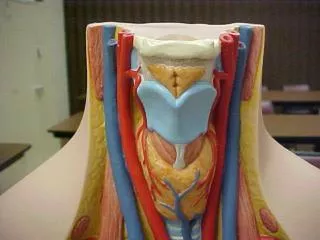
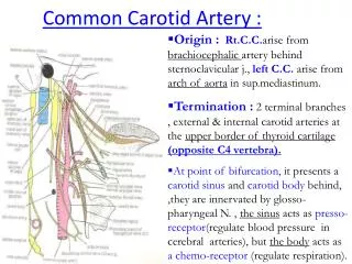
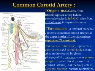
Carotid artery anatomy MCA Common carotid artery ACA Internal carotid Ophthalmic artery. Aortic arch
Carotid artery stenosis • The symptoms and pathologic substrate of carotid artery atherosclerotic occlusive disease were first described by C Miller Fisher in 1951 . • He related atherosclerotic disease at the carotid bifurcation to ischemic symptoms in the ipsilateral eye and brain.
Transient Ischemic attack • TIA has traditionally been defined as a focal, transient, neurological deficit of ischemic origin lasting less than 24 hours. • ‘‘brief episode of neurological dysfunction caused by a focal brain or retinal ischemia, with clinical symptoms typically lasting less than 1 hour and without evidence of [radiographically defined] infarction’’
Transient ischemic attacks • TIA is an important indicator of subsequent stroke risk with 25% to 30% of patients having a stroke over the ensuing 5 years. • the symptoms are related to the vascular territories involved. Most often, ischemic event related to carotid artery stenosis will produce symptoms referable to the middle cerebral artery territory, although the anterior cerebral artery can also be involved.
Transient ischemic attacks • Amaurosisfugax refers to transient monocular blindness caused by a small embolus to the ophthalmic artery. Intravascular plaquesmay sometimes be observed in retinal arterioles of patients experiencing amaurosisfugax (ie, Hollenhorst plaques).
Total carotid artery occlusion • When the internal carotid artery occludes completely, it can also cause low flow or embolic ischemic events depending upon the adequacy of collateral flow through the orbit and across the circle of Willis. • The greatest risk of low flow TIA or stroke is at the time of occlusion; the risk diminishes after the first year.
CLINICAL MANIFESTATIONS • Carotid bruit — An important sign of carotid stenosis heard over the site of the stenosis. • However, a carotid bruit in asymptomatic patients is a poor predictor for the presence of an underlying carotid stenosis and for the subsequent development of stroke.
Ischemic symptoms • Features of ocular ischemia or infarction include partial or complete blindness in one eye and an absent pupillary light respocerebralnse. • Hemispheric signs of infarction from carotid disease include contralateral homonymous hemianopsia, hemiparesis, and hemisensory loss. Specific signs of left hemisphere ischemia include aphasia, while right hemisphere ischemia may be manifest by left visuospatial neglect, constructional apraxia.
Ischemic symptoms • Atypical symptoms of internal carotid artery stenosis include unilateral limb shaking and transient loss of monocular vision upon exposure to bright light Syncope may be a rare consequence of bilateral carotid occlusive disease.
Ischemic symptoms • None of the above symptoms and signs is specific to carotid stenosis. As an example, temporal arteritis may produce ocular symptoms that are similar to those produced by carotid stenosis and should be considered in the differential diagnosis.
Evaluation of carotid artery stenosis History $ examination Sign & symptom of carotid artery territories ischemia YES NO Symptomatic carotid artery stenosis asymptomatic carotid artery stenosis In the large clinical trials addressing the management of carotid artery stenosis, the detection of "silent" infarcts on CT or MRI did not qualify the stenosis as symptomatic. In clinical practice, however, radiographic evidence of ischemia in the territory of a stenotic internal carotid artery may affect management.
CONVENTIONAL CEREBRAL ANGIOGRAPHY • Gold standred. • evaluation of the entire carotid artery system, providing information, plaque morphology, and collateral circulation which may affect management . • K • lllkk • The disadvantages of angiography include its invasive nature. • high cost, and risk of morbidity and mortality.
Carotid duplex ultrasound • K • lllkk • The absence of flow in the internal carotid artery may be due to occlusion, but hairline residual lumens can be missed on CDUS . • In addition, several studies have found that CDUS tends to overestimate the degree of stenosis. • the accuracy of CDUS relies heavily upon the experience and expertise of the ultrasonographer. • noninvasive, safe, and relatively inexpensive technique for evaluation the carotid arteries.
CT ANGIOGRAPHY • Compared with carotid duplex ultrasound, MRA is less operator dependent and does produce an image of the artery. However, MRA is more expensive and time-consuming than carotid duplex ultrasound and is less readily available. Furthermore, MRA may not be performed if the patient is critically ill, unable to lie supine, or has claustrophobia, a pacemaker or ferromagnetic implants • CT ANGIOGRAPHY — provides an anatomic description of the carotid artery lumen and allows imaging of adjacent soft tissue and bony structures. Three dimensional reconstruction allows relatively accurate measurements of residual lumen diameter. CTA may be particularly useful when CDUS is not reliable . • CTA -requires a contrast bolus comparable to that administered during a conventional angiogram. As a result, impaired renal function is a relative contraindication for its use, particularly in patients with diabetes or congestive heart failure.
MR ANGIOGRAPHY • MRA produces a reproducible three dimensional image of the carotid bifurcation with good sensitivity for detecting high grade carotid stenosis. • appear to be less accurate for detecting moderate stenosis.
MR ANGIOGRAPHY • Compared with carotid duplex ultrasound, MRA is less operator dependent and does produce an image of the artery. • However, MRA is more expensive and time-consuming than carotid duplex ultrasound and is less readily available. • Furthermore, MRA may not be performed if the patient is critically ill, unable to lie supine, or has claustrophobia, a pacemaker .
CHOICE OF IMAGING TEST • with suspected carotid stenosis first perform carotid duplex ultrasound. • Those with stenosis <50 percent are followed with serial examinations to determine if there is progression. • Those with stenosis ≥50 percent are evaluated with transcranial Doppler examination and MRA.
CHOICE OF IMAGING TEST • CTA is performed in lieu of MRA if there is a contraindication to MRA imaging and in cases where the duplex US and MRA do not agree. • Conventional angiography is rarely performed; indications include patients who cannot tolerate an MRA and in whom the risk of dye is sufficient to warrant bypassing CTA in favor of the gold standard examination. • Angiography is also done if nonatherosclerotic disease is suspected (eg, dissection, vasculitis).
Natural History • The risk of stroke over 5 years among patients with asymptomatic stenosiswas: • 8% for patients with less than 60% stenosis. • 14.8% for those with 60% to 74% stenosis. • 18.5% for those with 75% to 94% stenosis. • 14.7% for those with 95% to 99% stenosis. • In patients with symptomatic stenosis, the risk for recurrent stroke on medical therapy is 5% to 7% per year for all stroke.
Mangement MEDICAL • Optimization of preventive measure: • ASA. • Lipid lowering agent. • BP control. Surgical • Carotid endarterctomy. • Angioplasty and stenting. Which to choose ?????
Carotid endarterctomy • High risk procedure: • Bleeding. • Srtoke. • Nerve injury. • Stroke.
Carotid endarterectomy • CEA in asymptomatic patients should be considered a long-term investment. • CEA is suggested for medically stable men between the ages of 40 and 75 years with asymptomatic carotid stenosis of 60 to 99 percent who have a life expectancy of at least five years, provided the perioperative risk of stroke and death for the surgeon or center is less than 3 percent .
Carotidendarterectomy • Despite the increasing use of medical therapies for patients in ACST, the investigators concluded that it was doubtful that still wider use of better medical therapies would reduce the incidence of stroke much below the rate of 2 percent per year found in those allocated to deferral of CEA.
Carotid endarterectomy • CEA is recommended for patients with recently symptomatic carotid stenosis of 70 to 99 percent who have a life expectancy of at least five years, provided that the perioperative risk of stroke and death for the surgeon or center is less than 6 percent .
Carotid endarterectomy • CEA is not beneficial for symptomatic carotid stenosis of 30 to 49 percent, and CEA is harmful for symptomatic patients with less than 30 percent stenosis. • medical management is recommended rather than CEA for patients with symptomatic carotid stenosis that is less than 50 percent .