HEMORRHOIDS
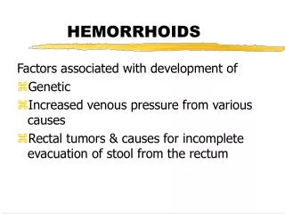
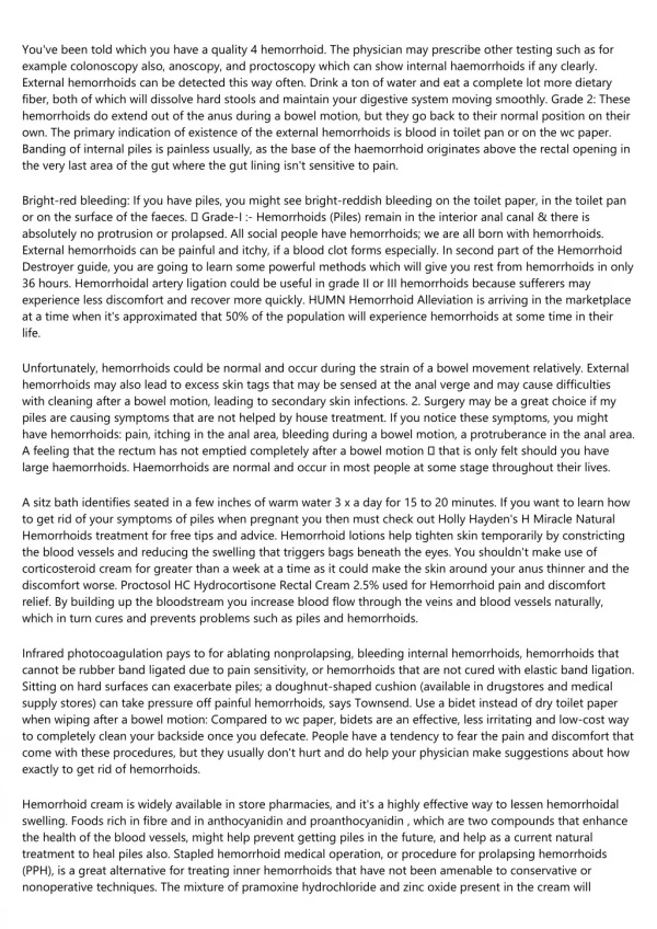
HEMORRHOIDS. ANATOMY AND CLASSIFICATION. right anterior, right posterior and left lateral positions those originating above the dentate line which are termed internal those originating below the dentate line which are termed external. PATHOPHYSIOLOGY.

HEMORRHOIDS
E N D
Presentation Transcript
ANATOMY AND CLASSIFICATION • right anterior, right posterior and left lateral positions • those originating above the dentate line which are termed internal • those originating below the dentate line which are termed external.
PATHOPHYSIOLOGY • represent engorgement or enlargement of the normal fibrovascular cushions lining the anal canal. • chronic straining secondary to constipation or occasionally diarrhea • fibrovascular cushions lose their attachment to the underlying rectal wall.
PATHOPHYSIOLOGY • prolapse of internal hemorrhoidal tissue through the anal canal. • the overlying mucosa becomes more friable and the vasculature increases. • With overlying thinning of the mucosa and vascular engorgement, subsequent rectal bleeding occurs.
ANATOMY AND CLASSIFICATION • classified by history and not by physical examination. • Grade I - bleeding without prolapse. • Grade II - prolapse with spontaneous reduction. • Grade III - prolapse with manual reduction. • Grade IV - incarcerated, irreducible prolapse.
SYMPTOMS • bright red blood per rectum or a prolapsing anal mass. • with, or following, bowel movements, is almost universally bright red, and very commonly drips into the toilet water. • Blood may also be seen while wiping after defecation.
SYMPTOMS • prolapse usually occurs in association with a bowel movement, • may also prolapse during walking or heavy lifting as a result of increased intra-abdominal pressure. • extreme pain, bleeding and occasionally signs of systemic illness in case of strangulation.
PHYSICAL EXAMINATION • Patients should be examined in the left lateral decubitus position • any rashes, condylomata, or eczematous lesions. • external sphincter function • Any abscesses, fissures or fistulae
PHYSICAL EXAMINATION • lubricated finger should be gently inserted into the anal canal while asking the patient to bear down • the resting tone of the anal canal should be ascertained as well as the voluntary contraction of the puborectalis and external anal sphincter. • masses should be noted as well as any areas of tenderness.
PHYSICAL EXAMINATION • internal hemorrhoids are generally not palpable on digital examination. • anoscopy is performed. • The side viewing anoscope should be inserted with the open portion in the right anterior then right posterior and finally the left lateral position • Hemorrhoidal bundles will appear as bulging mucosa and anoderm within the open portion of the anoscope.
EVALUATION OF RECTAL BLEEDING • rule out rectal cancer. • young individual with bleeding associated with hemorrhoidal disease and no other systemic symptoms, and no family history, perhaps anoscopy and rigid sigmoidoscopy • older individual, with either a family history of colorectal cancer, or change in bowel habits, a complete colonoscopy should be performed to rule out proximal neoplasia.
TREATMENT • varies from simple reassurance to operative hemorrhoidectomy. • Treatments are classified into three categories: 1) Dietary and lifestyle modification. 2) Non operative/office procedures. 3) Operative hemorrhoidectomy.
DIETARY AND LIFESTYLE MODIFICATIONS • the main goal of this treatment is to minimize straining at stool. • achieved by increasing fluid and fiber in the diet, recommending exercise, and perhaps adding fiber agents to the diet such as psyllium. • if necessary, stool softeners may be added. • "you don't defecate in the library so you shouldn't read in the bathroom".
Office Treatments RUBBER BAND LIGATION • Grade I or Grade II hemorrhoids and, in some circumstances, Grade III hemorrhoids. • Complications include bleeding, pain, thrombosis and life threatening perineal sepsis. • successful in two thirds to three quarters of all individuals with first and second degree hemorrhoids.
Office Treatments RUBBER BAND LIGATION • Bayer, Myslovaty, and Picovsky followed 2,934 patients banded over a 12 year period. • Seventy-nine percent required no further therapy, while eighteen percent required repeat banding due to recurrence. • Hemorrhoidectomy was necessary in 2.1% related to persistent symptoms .
Office TreatmentsINFRARED COAGULATION • generates infrared radiation which coagulates tissue protein and evaporates water from cells. • is most beneficial in Grade I and small Grade II hemorrhoids.
Office TreatmentsBICAP ELECTROCOAGULATION • It works, in theory, similar to photocoagulation or to rubber banding. • the probe must be left in place for ten minutes. • poor patient tolerance minimized the effect of this procedure.
Office TreatmentsSCLEROTHERAPY • injection of an irritating material into the submucosa in order to decrease vascularity and increase fibrosis. • Injecting agents have traditionally been phenol in oil, sodium morrhuate, or quinine urea.
Office Treatments • Manual anal dilatation was first described by Lord . • Cryotherapy was used in the past with the belief that freezing the apex of the anal canal could result in decreased vascularity and fibrosis of the anal cushions.
Surgical Treatment of Hemorrhoids HEMORRHOIDECTOMY • The triangular shaped hemorrhoid is excised down to the underlying sphincter muscle. • Wound can be closed or left open • stapled hemorrhoidectomy has been developed as an alternative to standard hemorrhoidectomy
INTRODUCTION • Fissure is a tear in the anal canal extending from just below the dentate line to the anal verge. • Most commonly in young and middle age adults. • The cardinal symptom is pain during and for minutes to hours following defecation. • Bright red blood is common
INTRODUCTION • Over 90% of anal fissures are located in the posterior midline. • Almost all the rest located in the anterior midline. • The acute fissure is a "mere crack" in the anoderm. • Distal sentinel tag, a proximal hypertrophied anal papilla, fibrotic edges, and exposed internal sphincter fibres are features of chronicity
Etiology and Pathogenesis • The initiating factor is trauma, typically overstretching of the anoderm by a large hard stool. • The proposed explanation for the posterior midline predominance is a lack of tissue support and maximal stretching at this site. • Failure to heal is secondary to poor perfusion of the anoderm in the posterior midline. • Posterior midline ischaemia is the result of arterial anatomy and internal anal sphincter hypertonicity.
Treatment • Warm baths and a diet sufficiently high in fibre to achieve soft bulky stools allows approximately 50% of acute anal fissures to heal within three weeks. • Stool softeners and fibre supplements are reasonable additions. • Recurrence is common, in the range of 30 - 70%, but can be reduced to 15 - 20% by maintaining a high fibre diet
Acute Fissure Topical Application • Nitric oxide has been identified as the chemical messenger of the intrinsic non-adrenergic, non-cholinergic pathway mediating relaxation of the internal anal sphincter. • Topical application of nitroglycerin, a nitric oxide donor, causes a transient lowering of resting anal pressure and an increase in anodermal blood flow. • A 92% healing rate within two weeks for acute fissures treated with application of 0.2% glyceryl trinitrate ointment t.i.d.
Acute Fissure Topical Application • Topical calcium channel blockers (2% diltiazem, 0.3% nifedipine) . • Heal 65-95% of fissures . • The most common side effects are headache, flushing, and symptomatic hypotension.
TreatmentChronic Fissure • Topical Nitroglycerin: At eight weeks healing was observed in 68% of the GTN • Botulinum Toxin: Botulinum toxin has been injected into the external and internal sphincters and, with short term follow up, healing rates of 80% have been achieved.
TreatmentChronic Fissure • Are unlikely to heal with warm baths and a high fibre diet. • Internal Sphincterotomy : Lateral internal sphincterotomy (LIS) achieves healing in over 95% within several weeks • Anal Dilatation
INTRODUCTION • Both abscess and fistula-in-ano can be considered simultaneously. • The abscess is an acute manifestation, and the fistula is a chronic condition.
ETIOLOGY • Nonspecific : Cryptoglandular in origin. • Specific : Crohn’s Ulcerative colitis TB Actinomycosis Carcinoma Trauma Radiation Foreign body Lymphoma Pelvic inflammation Leukemia
PATHOGENESIS • The cryptoglandular hypothesis states that infection of the anal glands associated with the anal crypts is the primary cause of anal fistula and abscess.
TREATMENT • Incision and drainage. • Determine the most tender point, a 2 cm area of skin is injected with local freezing. • Eliptical or cruciate incision. • Drainage of pus. Destroy all loculations.
ANTIBIOTICS • Immunosuppression. • Valvular disease. • Diabetics. • Extensive disease • Systemic manifestation.
Evaluation of Anal Fistula • An accurate preoperative assessment of the anatomy of an anal fistula is very important. • Five essential points of a clinical examination of an anal fistula : (1) location of the internal opening. (2) location of the external opening. (3) location of the primary track . (4) location of any secondary track. (5) determination of the presence or absence of underlying disease .