Download

1 / 18
240 likes | 851 Vues
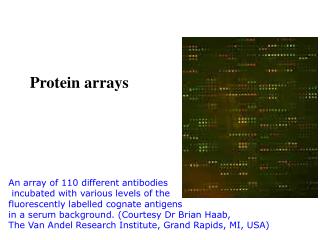
Protein Arrays. By: Nicole Therrien. Overview. What Are Protein Arrays? General Scheme Types of Arrays Analytical In-depth Example Functional Overview Example Reverse Phase Overview Example. What Are Protein Arrays?. Similar to DNA microarrays Plate, Probe, Attachment Advantage

E N D
Protein Arrays By: Nicole Therrien
Overview • What Are Protein Arrays? • General Scheme • Types of Arrays • Analytical • In-depth • Example • Functional • Overview • Example • Reverse Phase • Overview • Example
What Are Protein Arrays? • Similar to DNA microarrays • Plate, Probe, Attachment • Advantage • Poor correlation between mRNA and protein expression • Study protein interactions • Protein-Protein • Protein-Ligand • Protein-DNA • Monitor Disease States • Clinical Diagnostics
Types of Arrays • Analytical Microarrays • Functional Microarrays • Reverse Phase Microarrays
Analytical Microarrays • Profiles Mixture of Proteins • Measure Binding Affinity • Specificity • Protein Expression Levels • Most Common • 3 main probe types • Antibodies • Affibodies • Aptamers
Plate Set Up • Choose plate surface • Glass, Silicon • Attachment Method • Random Attachment • Covalent attachment by amines • Aldehyde • Epoxy • Adsorption • Nitrocellulose • Poly-L-Lysine • Acrylamide Gel Pads • Uniform Attachment • Affinity Tag • Nickel Coat & His tag • Streptavidin& Biotin • Spots vs. Wells • Sample incubated on plate with probes
Analytical Microarray Plates • Antibodies • 150 kDa • Standard • Affibodies • non-immunoglobulin-based affinity reagents • Based on Staphylococcus aureus protein A • Alpha-helices • No Disulfide • 6 kDa • Randomization of 13 AA in binding domain
Plates continued • Aptamers • Nucleic Acids • DNA, RNA, etc. • Peptides • Variable loop (10-20 AA) • Protein Scaffold • Bind Protein • Van der Waals Forces, H bonding, Electrostatic Interaction • Highly Specific • Engineered completely in test tube • In vitro selection
Sample Preparation • Sample extracted from cells or tissues • Bio-Rad assay • Labeled • Fluorescent Dye • Cy3/Cy5 via Lysines • Photochemical • Radioisotope • May interfere
Unlabeled • Antibody Sandwich • 2nd antibody with label incubated on top of sample • Surface Plasmon resonance • Measure electromagnetic waves • Angle changes in the order of 0.1° with 1 nm film adsorption • Needs special equipment • Don’t affect protein structure
Detection & Quantification • Scanner • Detects dye • Adjusts for background • Reference spots • Labeled known concentrations • Computational Analysis
Analytical Microarray Example • Individualized Medicine • Aptamers that recognize drug-resistant HIV-1 reverse transcriptase • In vitro selection • Attached with neutravidin & biotin • Incubated w/ M3 or WT HIV-1 RT • 1° Antibody • Rabbit Anti-HIV-1 • 2°Antibody • Goat Anti-rabbit Cy-3 label • Read by microarray scanner
Functional Microarrays • Plates • Full length proteins & protein domains • Functional • Samples • Purified & Labeled • Nucleic Acids • Proteins • Lipids • Small Molecules
Functional Array Example • Protein-Small Molecule Interaction • Plate has whole proteome • Monitor Specificity • Off-target effects
Reverse Phase Microarrays • Plates • Cell Lysate • Sample • Antibodies of interest • Primary • Attach to spots • Secondary • Attach to primary • Labeled • Detect Altered Proteins • Post-translation modification problems • Disease
Reverse Phase Example • Quantitative cell signaling analysis reveals down-regulation of MAPK pathway activation in colorectal cancer • Mitogen-activated protein kinases • Role in colonic cancer • Lysates of WT and Cancerous cells attached via nitrocellulose • Phospho-specific Rabbit antibodies • Staining • Ras mutations in colorectal cancer • Thought to increase MAPK pathway • Cautioned against kinase inhibition therapy
References • Amaratunga, Dhammika, and Javier Cabrera. Exploration and Analysis of DNA Microarray and Protein Array Data. Hoboken, NJ: John Wiley, 2004. • Gullman, Christian, et al. "Quantitative Cell Signalling Analysis Reveals Down-Regulation of MAPK Pathway Activation in Colorectal Cancer." Journal of Pathology 218 (2009): 514-19 • Hall, David et al.. "Protein Microarray Technology." Mechanisms of Ageing and Development 128.1 (2007): 161-67. \ • Li, Na et al. "Aptamers That Recognize Drug-Resistant HIV-1 Reverse Transcriptase." Nucleic Acids Research 36.21 (2008): 6739-751. • "Panorama Antibody Array Frequently Asked Questions." Sigma Aldrich Home. Web. 10 Apr. 2010. <http://www.sigmaaldrich.com/life-science/cell-biology/protein-arrays/protein-arrays-applications/faq.html>. • "Surface Plasmon Resonance - Wikipedia, the Free Encyclopedia." Main Page - Wikipedia, the Free Encyclopedia. Web. 10 Apr. 2010. <http://en.wikipedia.org/wiki/Surface_plasmon_resonance>. • Tao, Sheng-Ce, et al. "Applications of Protein Microarray Technology." Combinatorial Chemistry & High Throughput Screening 10 (2007): 706-18.