Download

1 / 22
230 likes | 700 Vues
Adrenals, Lymphnodes, Gall Bladder, and Pancreas. Jane MacLellan. Adrenal Glands. In the dog - peanut shaped In the cat - more oval Located cranial and medial to kidneys Left - caudal to the branches of the aorta Right - adjacent to caudal vena cava

E N D
Adrenals, Lymphnodes, Gall Bladder, and Pancreas Jane MacLellan
Adrenal Glands • In the dog - peanut shaped • In the cat - more oval • Located cranial and medial to kidneys • Left - caudal to the branches of the aorta • Right - adjacent to caudal vena cava • Locate kidney, then fan medially with probe • Hypoechoic - similar to blood vessels • Can be hard to distinguish - use doppler • Overlying bowel can obscure
Adrenal Gland Disease • Measure length and width • Length with vary between animals • Proportional to body weight • Width does not • Width may increase with disease • Normal width = < 0.74 cm • Note: New paper suggests dogs < 10Kg normal width < 0.6 cm
Adrenal Gland Disease • Pituitary dependent hyperadrenocorticism • Bilaterally enlarged • Normal shape - but ‘plump’ • Thickened poles • Uniformly hypoechoic • Nodular hyperplasia • Normal size does not r/o PDH
Adrenal Gland Disease • Adrenal tumor • Gland enlargement • Abnormal shape • Change in echotexture • Unilateral masses more common • Can’t distinguish benign from malignant tumors • May be able to tell if invading surrounding tissue
Lymph nodes • More sensitive then radiographs • Medial iliac and jejunal lymph nodes • Large • More often seen when normal • Normal - same echogenicity as surrounding mesentery • Easier to see in young, thin animals • When enlarged, more hypoechoic • Can do ultrasound guided FNA
Lymph nodes • Medial iliac • Near terminal portion of aorta and caudal vena cava • Not normally seen unless enlarged • Bladder, prostatic neoplasia • Visceral • Seen when doing routine scan
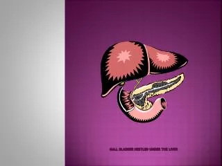
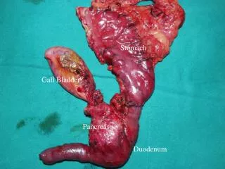
Gall Bladder • Visualized just right of midline in liver • Size is variable - depending on last meal • Fasting or anorexia • In cats, can be bi-lobed • Things you might see • Thickened wall • Stones • Mucoceles • Cholestasis • Cholecystitis • “Sludge” • Icterus
Thickened Wall • Wall normally thin, echogenic, poorly visualized • <1mm in cats, slightly thicker in dog • Double layered - inside and outside surfaces – Halo sign • Thickening is a non-specific sign • Chronic hepatitis • Cholecystitis • Cholangiohepatitis • Right CHF • Hypoalbuminemia • Sepsis • Neoplasia
Sludge • Commonly seen • Especially if haven’t eaten recently • Dependent
Mucocele • Cystic mucinous hyperplasia • Proliferation of GB epithelium • Increased mucin production • Marked distension of the GB • Kiwi appearance
Choleliths • Uncommon • Incidental finding • Should be noted - cholecystitis or biliary obstruction • Hyperechoic • Acoustic shadowing • Mobile
Bile Duct • Seen as a continuation of the GB • Dogs - not consistently seen • Should be < 3mm • Cats - more often seen • Should be < 4mm • Ventral to portal vein • Extrahepatic obstruction • Dilation of GB and bile duct
GB Artifacts • Mirror image duplicate • Sound wave bounces off the diaphragm, echos off the gall bladder back towards diaphragm, reflected towards the transducer • Refraction • When sound waves go through tissues of different acoustic impedance • Acoustic enhancement • Less attenuation compared to liver
Pancreas • Normal is routinely difficult to visualize • Echogenicity similar to surrounding fat • No defined capsule • Less echogenic then spleen, more echogenic then liver • Right limb just dorsal to duodenum • More likely to see in puppies, thin dogs, or with free abdominal fluid
Pancreatitis • Acute - surrounded by a hyperechoic area • Due to peri-pancreatic fat necrosis • Severe - mixed echogenicity • Chronic - hyperechoic pancreas • Due to pancreatic fat necrosis • Pancreatic pseudocysts • Mass effect
Pancreas • Neoplasia • Difficult to identify • Looks similar to pancreatitis (mass effect) • Fluid accumulation • Invasion of surrounding tissues • Evidence of metastasis in other organs