Download
1 / 44
440 likes | 785 Vues
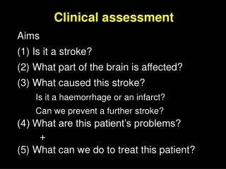
Clinical assessment. Aims (1) Is it a stroke? (MSD) (2) What part of the brain is affected? (3) What caused this stroke? Is it a haemorrhage or an infarct? Can we prevent a further stroke? (4) What are this patient’s problems? + (5) What can we do to treat this patient? (RIL).

E N D
Clinical assessment Aims (1) Is it a stroke? (MSD) (2) What part of the brain is affected? (3) What caused this stroke? Is it a haemorrhage or an infarct? Can we prevent a further stroke? (4) What are this patient’s problems? + (5) What can we do to treat this patient? (RIL)
Is it a stroke? (a) The setting (or demographics) • age • hypertension • smoking • diabetes • cholesterol • presence of other vascular disease (b) The nature of the event • onset • course • focal vs general symptoms • “negative” symptoms (loss of function) • associated symptoms
Stroke mimics • Migraine • Epilepsy • Structural brain lesions • SDH, Tumour, abscess • Metabolic/toxic disorders • hypoglycemia • Vestibular disorders • Psychological disorders • Demyelination • Mononeuropathy
1. Confirms the diagnosis of stroke 2. Allows better selection of imaging 3. Gives an indication of cause 4. Gives an indication of prognosis Localisation: Why bother?
Localising the lesion depends on a basic understanding of neuroanatomy • the cortex • the homunculus • deep white matter • the brainstem • the vascular supply
What part of the brain is affected? • Left or right • Carotid territory or vertebrobasilar territory • Cerebral hemispheres or brainstem • Cortex or deep white matter
Neuroanatomy 1: Left or Right? • Crossing of sensory and motor fibres • corticospinal tracts - lower medulla • spinothalamic fibres - spinal cord • dorsal columns - upper medulla • Cerebellar lesions result in ipsilateral deficits • The “dominant hemisphere” • Language function localises to left hemisphere • Awareness of body localises to right hemisphere • Visual pathways • monocular vs homonymous deficits
A small stroke there (or there) will result in a major deficit as the fibres are packed close together Neuroanatomy 4: deep white matter
Cranial nerve signs suggest localisation to (and within) the brainstem Neuroanatomy 5: the brainstem
The carotid system supplies most of the hemispheres and cortical deep white matter The vertebro-basilar system supplies the brain stem, cerebellum and occipital lobes Neuroanatomy 6: the vascular supply
So, from the symptoms and signs you observe, you can tell: • what side of the brain is affected • whether the lesion is in the brainstem (a brainstem stroke) • whether the cortex is involved (a cortical stroke) • or if the lesion is in the deep white matter (a lacunar stroke) • what blood vessel is involved
Is it a stroke? • Male, 58 years • Headache for 4 weeks • 10 days of gradually increasing right side weakness • O/E: • poor concentration • slow speech, unable to follow commands • right face & arm weak, walking OK • papilloedema
68 year old woman • On warfarin for AF • Previous mild stroke • Sudden onset left leg weakness • O/E: • unaware of problems • dense weakness of left, loss of sensation • doesn’t look to left • mildly drowsy • INR 2.9
75 year old man • Hypertension, diabetes mellitus • sudden onset dizziness & vomiting, unable to walk • O/E: • constricted pupil on left • nystagmus in all directions • ataxia of left arm & leg • loss of PP on right
69 year old woman • hypertension, smoker • 2 days ago episode of right arm & leg weakness • sudden onset worse right sided weakness • O/E: • slurred speech only • equal weakness of face, arm and leg; unable to walk; sensation OK • alert
The pathology 2 processes result in a stroke: (1) Infarction • 85% of strokes • occlusion of a vessel by thrombosis or embolus (2) Haemorrhage • 15% of strokes • rupture of a vessel results in bleeding into the substance of the brain
Intracerebral Haemorrhage • Usually caused by hypertension • thickening & weakening of walls of small arteries/arterioles • formation of small aneurysms • rupture produces a large blood filled cavity that acts as a SOL • typically basal ganglia or thalamus
Cerebral Infarction • Infarction is caused by failure of blood flow to a region • damage to the brain is due to: • ischaemia • oedema surrounding the ischaemic area • sources of occlusion of vessels: • thrombosis of small vessels - hypertensive lipohyalinosis - lacunar infarcts • thrombosis of larger vessels • embolus from extracranial vessels or heart
Thrombo-embolism • At least 1/3 of strokes are due to emboli from heart or ICA • small clot breaks off from a larger thrombus • it becomes lodged in a distal smaller vessel, producing an infarct • Cardiac sources of embolus are common with conditions such as AF or prosthetic valves
Cerebral Infarction A recent infarct in the right temporal lobe - loss of gray-white margin, swelling Old lacunar infarct of right putamen & internal capsule Old infarct of the right MCA - cystic formation & enlargement of the ventricle
Haemorrhagic infarction • Usually infarcts are bland - necrosis only • Occasionally there is haemorrhage seen in the infarct • occurs in embolic infarcts • due to spontaneous lysis of the clot reperfusion of damaged vessels • often asymptomatic The bleeding is petichial and confined to the cortex
Features of an infarct depend on the blood vessel occluded 3 main cortical vessels: ACA, MCA, PCA
Distinguishing haemorrhage from infarct clinically is difficult & unreliable • On history: • severe headache • vomiting within 2 hours of onset • On examination: • marked hypertension • altered conscious state • Increasing evidence to suggest that mild events may be due to PICH • Scanning is the only acceptable method
Brain Imaging • Rationale: • to exclude (rare) stroke mimics eg SDH • to distinguish between haemorrhage and infarct • Plain CT is the imaging technique of choice • available, rapid • reliably differentiates haemorrhage: blood is white
Intracerebral haemorrhage on CT • Is always seen • apparent immediately • lasts 1 week • then disappears and looks like an infarct
Ischaemic stroke on CT • Infarcts seen as areas of hypodensity • become more obvious as time progresses • small infarcts appear later than large ones • overall, 40% strokes have normal CT • posterior fossa difficult
Haemorrhagic Transformation Haemorrhage seen at the margins of an infarct
MR in acute stroke • Advantages: • much better at defining the anatomy • shows ischaemic changes earlier, and in a greater proportion of patients • diffusion weighted imaging can show ischaemia within minutes-hours, and differentiate between old and new lesions • MRA allows imaging of blood vessels non-invasively • Disadvantages: • expense, time, lack of access to the patient
CT brain (3 hours) ? R MCA hypodensity DWI (24 hrs) obvious R MCA infarct MRA (24 hrs) dissection R ICA with distal occlusion MRI in acute stroke: an example A 42 year old man with headache and left hemiparesis
What caused this infarct? • The clinical assessment may provide clues to the likely cause • history - demographics atheroma • examination - carotid bruits atheroembolism, heart abnormalities (AF, murmurs) cardioembolism • Localisation provides the best clues: • cortical stroke cardiac or large artery embolus • lacunar stroke small vessel disease • brainstem stroke local atheroma
Knowing the likely cause tells you how to investigate further... • If cortical stroke: • look closely at the heart (ECG, ?Echo) • look for carotid atheroma (Carotid duplex) • specialised tests if young • If lacunar stroke: • look closely for risk factors, fewer tests
What caused this haemorrhage? • According to age: <45 years AVM 45-69 years small vessel disease >70 years cerebral amyloid small vessel disease • According to location: Lobar amyloid, AVM, small vessel Deep white small vessel disease
PICH - two types Lobar bleed (from cerebral amyloid) Basal ganglia bleed (from right caudate nucleus)
Prognosis after Intracerebral Haemorrhage • 40% dead in first 7 days • 50% dead in first 30 days • 62% dead by 1 year more likely to die early, but mortality reduces thereafter of the 40% alive, 30% are independent
Prognosis after cerebral infarction • For all: 5% dead by 7 days 10% dead by 1 month 23% dead by 1 year • For large cortical strokes: 60% dead, 35% disabled • For lacunar strokes: 11% dead, 26% disabled
Clinical assessment Aims (1) Is it a stroke? (MSD) (2) What part of the brain is affected? (PJH) (3) What caused this stroke? (PJH) Is it a haemorrhage or an infarct? Can we prevent a further stroke? (4) What are this patient’s problems? + (5) What can we do to treat this patient? (RIL)