Download

1 / 84
1.14k likes | 2.89k Vues
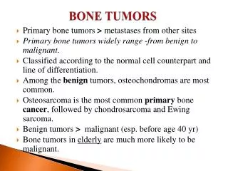
Bone Tumors and Tumor-like Conditions. Prof. Mamoun Kremli AlMaarefa College. Objectives. Bone tumors: Primary: Benign – Malignant Secondaries in bone Tumor-like conditions Bone cysts How to read x-ray of a bone lesion. Classification – predominant tissue.

E N D
Bone Tumors andTumor-like Conditions Prof. Mamoun Kremli AlMaarefa College
Objectives • Bone tumors: • Primary: • Benign – Malignant • Secondaries in bone • Tumor-like conditions • Bone cysts • How to read x-ray of a bone lesion
Clinical presentation - history • Prolonged history: • In most benign lesions • Some malignant: slow growing / in pelvis (expandable) • Age: • Childhood and adolescence • Most benign, and some malignant (e.g. Ewings sarcoma) • 4th – 5th decade: • Chondrosarcoma and fibrosarcoma • Sixth decade: • Myeloma (the commonest primary malignant bone tumor) • Over 70 yrs: • Metastatic lesions are the commonest
Clinical presentation - history • Pain: • In both malignant and benign • May be caused by: • Rapid expansion – stretching of tissues • Central hemorrhage or degeneration • Insipient pathological fracture • Tense encapsulation in bone (e.g. osteoid osteoma) • Swelling • H/O Trauma • Neurological symptoms • Pressure on nerve / stretching the nerve • Pathological fracture
Clinical examination • A mass (lump) • Location • Discrete or ill-defined • Tenderness • Warm • Pulsatile • Mobility • ….etc • Range of motion • LN, pelvis, abdomen, chest, spine
Imaging – x-rays • Which bone, and which site in bone? • Solitary or multiple? • Bone forming or bone eating? • Margins: well-defined or ill-defined? • Calcifications in the lesion? • Is cortex eroded or destroyed? • Is there periosteal new bone formation? • Soft tissue extension?
Location Orthopedic Radiolgy. A Greenspan. Lippincott-Raven
Radiographic features Orthopedic Radiolgy. A Greenspan. Lippincott-Raven
The Border Orthopedic Radiolgy. A Greenspan. Lippincott-Raven
The Matrix Orthopedic Radiolgy. A Greenspan. Lippincott-Raven
Type of Bone Destruction Orthopedic Radiolgy. A Greenspan. Lippincott-Raven
Type of Periosteal Reaction Orthopedic Radiolgy. A Greenspan. Lippincott-Raven
Soft Tissue Extension Orthopedic Radiolgy. A Greenspan. Lippincott-Raven
Benign Vs. malignant Orthopedic Radiolgy. A Greenspan. Lippincott-Raven
Other imaging • Bone scan (Tc99): • Shows the site of lesion / and skip lesions • CT: • Intraosseous and extraosseous structure and extension • Good in deep bones (pelvis, spine) • MRI: • Tumor spread • Within bone, into joints, into soft tissue • Relation to vessels • Soft tissue and cartilage tumors
Lab, investigations • Look for infection • Look fro metabolic disease (brown tumor) • Anemia, raised ESR • S. Alkaline phosphatase • Bence Jones protein in urine: myeloma • S. Acid phosphatase: prostatic carcinoma • Raised s Calcium in metastasis
Biopsy • Diagnostic • Needle biopsy: • CT- guided • Inthe line of further surgical incision • Representative sample • ? frozen section confirmation of a good sample • Open biopsy: • After all imaging techniques completed • More reliable – significant morbidity • Site considering further surgery • From boundaries • Excision biopsy for almost certainly benign tumors
Differential diagnosis • Soft tissue hamartomas • Myositis ossificans • Stress fracture: • Histopath. may be confused with osteosarcoma? • Tendon avulsion injuries • Near hip and knee (e.g. Osgood-Schlatter) • Infection • Gout: • Large gouty typhus • Other bone lesions: • Cortical defects, bone infarcts, “bone islands”
Staging • How does the tumor behave? • Aggressiveness • How far has it spread? • Extent
Tumor Excision • Intracpsular • Marginal • Wide local • Radical • Amputation Apley’s System of Orthop. And Fractures
Benign bone lesions • Non-ossifying fibroma • Fibrous dysplasia • Osteoid osteoma / osteoblastoma • Chondroma / chondroblastoma • Osteochondroma • Simple bone Cyst • Aneurysmal bone cyst • Giant cell tumor
Non-ossifying fibroma • Another name: • Fibrous cortical defect • The commonest benign lesion of bone • Asymptomatic • Incidentally discovered • Children: • Disappears later • Common site: • Metaphysis of long bones • Treatment: • Observation • Surgery if v large Apley’s System of Orthop. And Fractures Orthopedic Radiolgy. A Greenspan. Lippincott-Raven
Non-ossifying fibroma Non-ossifying fibroma ……..………Fibrous cortical defect Orthopedic Radiolgy. A Greenspan. Lippincott-Raven
Fibrous Dysplasia • Developmental disorder • Trabecular bone replaced by fibrous tissue • Types: • Monstatic • Monomelic • Polystatic • Site: • Prox. Femur: • Shepherd’s crook • Tibia, humerus. Ribs, cranio-facial • Deformity of bone Apley’s System of Orthop. And Fractures
Polystotic Fibrous Dysplasia Orthopedic Radiolgy. A Greenspan. Lippincott-Raven
Osteoid osteoma • Small tumor (<1 cm) • Young adults • Pain, pain, pain • Relieved by Salicylates • Sites: Femur, tibia, spine • X-ray: • Small radiolucent “nidus” • Surrounded by sclerotic bone • CT: Shows “nidus” better • Tc scan: hot • Treatment: surgical excision, or thermal ablation Apley’s System of Orthop. And Fractures
Osteoid osteoma • Small tumor (<1 cm) • Young adults • Pain, pain, pain • Relieved by Salicylates • Sites: Femur, tibia, spine • X-ray: • Small radiolucent “nidus” • Surrounded by sclerotic bone • CT: Shows “nidus” better • Tc scan: hot • Treatment: surgical excision, or thermal ablation Orthopedic Radiolgy. A Greenspan. Lippincott-Raven
Osteoid Osteoma • 7 year old boy
Osteoid Osteoma Orthopedic Radiolgy. A Greenspan. Lippincott-Raven
Osteoblastoma • A giant ostoid osteoma • Spine and flat bones Orthopedic Radiolgy. A Greenspan. Lippincott-Raven
Chondroma (Enchondroma) • Incidentally discovered • Young age • Tubular bones of hands and feet • X-ray: • Well-defined, central lesion • At junction of metaphysis with diaphysis • Flake of calcification are characteristic • Malignant transformation • Rare in solitary • 30% in multiple (Ollier’s disease) Orthopedic Radiolgy. A Greenspan. Lippincott-Raven
Chondroma (Enchondroma) • Incidentally discovered • Young age • Tubular bones of hands and feet • X-ray: • Well-defined, central lesion • At junction of metaphysis with diaphysis • Flake of calcification are characteristic • Malignant transformation • Rare in solitary • 30% in multiple (Ollier’s disease) Slide Atlas of Orthop Pathology, P Bullough. Gower Med P
Chondroma (Enchondroma) Orthopedic Radiolgy. A Greenspan. Lippincott-Raven
Enchondromatosis (Ollier’s) • Many lesions • Malignant transformation: 30% Orthopedic Radiolgy. A Greenspan. Lippincott-Raven
Enchondromatosis (Ollier’s) • Many lesions • Malignant transformation: 30% Orthopedic Radiolgy. A Greenspan. Lippincott-Raven
Chondroblastoma • In epiphysis • Proximal humerus, femur, tibia Apley’s System of Orthop. And Fractures Orthopedic Radiolgy. A Greenspan. Lippincott-Raven
Osteochndroma (Exostosis) • A common lesion • Ends of long bone • Bony overgrowth • Away from epiph. Late • Covered by cartilage • Growth: • Stops when epiphysis close • If continues later: • ? Malignant transformation Orthopedic Radiolgy. A Greenspan. Lippincott-Raven
Osteochndroma (Exostosis) Apley’s System of Orthop. And Fractures Orthopedic Radiolgy. A Greenspan. Lippincott-Raven
Multiple Exostosis • Many lesions • Causes growth disturbance Orthopedic Radiolgy. A Greenspan. Lippincott-Raven
Simple bone cyst • Solitary – unicameral • Children • Metaphysis • Prox. Humerus and Femur • Not a tumor • Not seen in adults • Heals spontaneously • Pathological fracture / incidental • Aspirate is clear straw-colored Orthopedic Radiolgy. A Greenspan. Lippincott-Raven
Simple bone cyst • Solitary – unicameral • Children • Metaphysis • Prox. Humerus and Femur • Not a tumor • Not seen in adults • Heals spontaneously • Pathological fracture / incidental • Aspirate is clear straw-colored Orthopedic Radiolgy. A Greenspan. Lippincott-Raven
Simple bone cyst • Treatment: • Small, reducing: leave alone • Increasing in size, active • Multiple bone marrow injections • Pathological fracture • Treat fracture • Cyst might heal • Recurrent / injection failed: • Surgical curettage and bone grafting Orthopedic Radiolgy. A Greenspan. Lippincott-Raven
Aneurysmal bone cyst • Young adults • Metaph. of long bone • X-ray: • Well-defined cyst • Trabeculated • Eccentrically placed • Ballooning • Bloody content Orthopedic Radiolgy. A Greenspan. Lippincott-Raven
Giant-Cell Tumor • Unknown origin • Giant cells abundant • Behavior: • One third benign • One third locally aggressive • One third (less) with distant metastasis • Young adults • Common sites: • Around knee • Proximal humerus • Distal radius Orthopedic Radiolgy. A Greenspan. Lippincott-Raven
Giant-Cell Tumor • Unknown origin • Giant cells abundant • Behavior: • One third benign • One third locally aggressive • One third (less) with distant metastasis • Young adults • Common sites: • Around knee • Proximal humerus • Distal radius Apley’s System of Orthop. And Fractures
Giant-Cell Tumor • Eccentric lesion • Radiolucent • Soap bubble • Abuts against the joint • Thin cortex • Margins may be clear / unclear • Depends on aggressiveness • Treatment • Curettage & bone grafting • More wide excision in recurrent and aggressive lesions Apley’s System of Orthop. And Fractures
Giant-Cell Tumor Apley’s System of Orthop. And Fractures