Download
1 / 82
820 likes | 1.11k Vues
ELEKTRONOVÉ A IONTOVÉ SPEKTROSKOPIE. kód Měření povrchových vlastností pevných látek. Doc. RNDr. Karel Mašek, Dr. Skupina fyziky povrchů KEVF. Spektroskopie obecně. Primární činidlo – rtg záření, elektrony, ultrafialové záření, synchrotronové záření, ionty, tepelná energie

E N D
ELEKTRONOVÉ A IONTOVÉ SPEKTROSKOPIE kód Měření povrchových vlastností pevných látek Doc. RNDr. Karel Mašek, Dr. Skupina fyziky povrchů KEVF
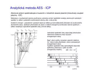
Spektroskopie obecně • Primární činidlo – rtg záření, elektrony, ultrafialové záření, synchrotronové záření, ionty, tepelná energie • Měříme – energetické rozdělení vyletujících (sekundárních) elektronů nebo iontů • Zjišťované informace – chemické složení, chemický stav, čistota, vazby (hloubka informace), reaktivita povrchu, struktura povrchu • SPEKTRUM = závislost intenzity na měřené energii nebo hmotnosti • Intenzita – většinou počet pulsů za vteřinu, proud
Schéma aparatury Schéma aparatury
Hlavní parametry spektroskopických metod • Typ informace – chemické složení, struktura, stav povrchu apod…. • Hloubka informace – záleží na průniku primárních částic do látky a únikové hloubce signálních částic z látky • Poškození analyzovaného vzorku • Citlivost a rozlišení dané metody
Elektronové spektroskopie • XPS (X-ray photoelectron spectroscopy) fotoelektronová spektroskopie • AES (Auger electron spectroscopy) Augerova elektronová spektroskopie • UPS (ultraviolet photoelectron spectroscopy) ultrafialová fotoelektronová spektroskopie • EELS (electron energy loss spectroscopy) spektroskopie charakteristických ztrát a modifikace (HREELS, RHEELS) • SRPES (synchrotron radiation photoelectron spectroscopy) fotoelektronová spektroskopie buzená synchrotronovým zářením
Iontové spektroskopie • SIMS (Secondary Ion Mass Spectroscopy) hmotnostní spektroskopie sekundárních iontů, typy SSIMS a DSIMS • ISS (Ion Scattering Spectroscopy) spektroskopie rozptýlených iontů • LEIS (Low Energy Ion Spectroscopy) spektroskopie nízkoenergetických iontů • TDS (Thermodesorption Spectroscopy) termodesorpční spektroskopie • TPR (Temperature Programmed Desorption) Teplotně programovaná reakce
method AES XPS UPS SSIMS DSIMS ISS RBS základní informace chemické složení chemické složení struktura val. pásu povrchové chemické vazby chemické složení (izotopy) chemické složení (izotopy) chemické složení chemické složení citlivost (det. limit) ppm 1000 1000 103 - 104 10 10-3 104 104 povrchová citlivost (hloubkové rozlišení) nm 1 1 0.3 0.6 10 0.3 10 laterální rozlišení 25 nm 0.1 mm 1 mm 1 mikron 50 nm 1 mm 1 mikron nedestruktivní? víceméně ano ano víceméně ne víceméně ano hloubkový profil v kombinaci s odprašováním, nebo změnou energie a úhlu dopadu v kombinaci s odprašováním, nebo změnou energie fotoelektronů a úhlu detekce - ano (pomalý) ano v kombinaci s odprašováním ano další informace valence, chemický stav valence, chemický stav, struktura (ARPES) vazebná geometrie (ARUPS) povrchové sloučeniny povrchové sloučeniny struktura (LEIS) struktura modifikace mapování a zobrazení prvků (SAM) zobrazení mikropóry Srovnání metod
Instrumentální vybavení Detektor Interface Primární zdroj Analyzátor • Primární zdroj • Rtg záření Al, Mg Kα • elektrony 50 – 5000 eV • UV záření He výboj • synchrotronové záření • 40 – 1000 eV • - Zdroj iontů 50 – 5000 eV Vstupní optika PC vzorek
4-mřížkový analyzátor LEED – difrakce nízkoenergetických elektronů AES – Augerova spektroskopie
- Vouter + Cylindrický analyzátor (CMA) Jednoduchý CMA Vnější válec Vnitřní válec se štěrbinami Vzorek Detektor (channeltron) apertura Koaxiální elektronovédělo
Cylindrický analyzátor (CMA) Dvojitý CMA (s brzdnýmpolem)
Hemisférický analyzátor HMA Elektronové spektroskopie – XPS, UPS, AES, EELS, SRPES Lepší rozlišení Citlivost závisí na velikosti sfér
Vouter dN(E)/dE Lock-in zesilovač DN(E) N(E) DE Energie Způsob měření • Přímé spektrum – proud nebo počet pulsů za jednotku času • Derivované spektrum – první derivace (někdy i druhá derivace) signálu, v případě analýzátoru s brzdným polem získáme přímé spektrum střídavá modulace (~1 V, 10 kHz)
Způsob měření Derivované spektrum Přímé spektrum
Způsob měření • Detektor • násobič • kanálek (channeltron) • pole kanálků • kanálková destička (channelplate) Lock-In Interface AC modulace Řídící jednotka, zdroj • Elektronika analyzátoru • potřebná řídící a napájecí napětí • komunikace s počítačem • Snímání signálu z detektoru PC • PC ainterface(převodníky, čítače, komunikační karty) • komunikace s řídící jednotkou analyzátoru • generování řídících příkazů nebo signálů • akumulace dat, jejich záznam a zobrazení
Způsob měření • Detektor • násobič • kanálek (channeltron) • pole kanálků • kanálková destička (channelplate) Detektorová jednotka Interface Řídící jednotka, zdroj • Elektronika analyzátoru • potřebná řídící a napájecí napětí • komunikace s počítačem • Snímání signálu z detektoru PC • PC ainterface(převodníky, čítače, komunikační karty) • komunikace s řídící jednotkou analyzátoru • generování řídících příkazů nebo signálů • akumulace dat, jejich záznam a zobrazení
Elektronové spektroskopie • Fotoelektronová spektroskopie – XPS, UPS, SRPES • Elektrony buzené spektroskopie – AES, EELS
M2 3s M1 2p3/2 L3 2p1/2 L2 2s L1 1s K L3 Fotoelektrický jev foton fotoelektron BE vazebná energie hν energie fotonu KE kinetická energie Ef energie konečného stavu Ei energie počátečního stavu
KE = hv – BE NOTE - the binding energies (BE) of energy levels in solids are conventionally measured with respect to the Fermi-level of the solid, rather than the vacuum level. This involves a small correction to the equation given above in order to account for the work function (φ) of the solid, but for the purposes of the discussion below this correction will be neglected.
XPS • For each and every element, there will be a characteristic binding energy associated with each core atomic orbital i.e. each element will give rise to a characteristic set of peaks in the photoelectron spectrum at kinetic energies determined by the photon energy and the respective binding energies. • The presence of peaks at particular energies therefore indicates the presence of a specific element in the sample under study - furthermore, the intensity of the peaks is related to the concentration of the element within the sampled region. Thus, the technique provides a quantitative analysis of the surface composition and is sometimes known by the alternative acronym , ESCA (Electron Spectroscopy for Chemical Analysis). • The most commonly employed x-ray sources are those giving rise to : Mg Kα radiation : hv = 1253.6 eV Al Kα radiation : hv = 1486.6 eV • The emitted photoelectrons will therefore have kinetic energies in the range of 0 - 1250 eV or 0 - 1480 eV Since such electrons have very short lifetimes in solids, the technique is necessarily surface sensitive.
The diagram below shows a real XPS spectrum obtained from a Pd metal sample using Mg Ka radiation the main peaks occur at kinetic energies of ca. 330, 690, 720, 910 and 920 eV.
Since the energy of the radiation is known it is a trivial matter to transform the spectrum so that it is plotted against BE as opposed to KE. The most intense peak is now seen to occur at a binding energy of ca. 335 eV
1. the valence band (4d,5s) emission occurs at a binding energy of ca. 0 - 8 eV ( measured with respect to the Fermi level, or alternatively at ca. 4 - 12 eV if measured with respect to the vacuum level ). 2. the emission from the 4p and 4s levels gives rise to very weak peaks at 54 and 88 eV respectively 3. the most intense peak at ca. 335 eV is due to emission from the 3d levels of the Pd atoms, whilst the 3p and 3s levels give rise to the peaks at ca. 534/561 eV and 673 eV respectively. 4. the remaining peak is not an XPS peak at all ! - it is an Auger peak arising from x-ray induced Auger emission. It occurs at a kinetic energy of ca. 330 eV (in this case it is really meaningless to refer to an associated binding energy).
Spin-Orbit Splitting Closer inspection of the spectrum shows that emission from some levels (most obviously 3p and 3d ) does not give rise to a single photoemission peak, but a closely spaced doublet. We can see this more clearly if, for example, we expand the spectrum in the region of the 3d emission ...
hn = Eb(k) + F + Ec E = hn - Eb(k) - Fs
Example 1 : Oxidation States of Titanium Titanium exhibits very large chemical shifts between different oxidation states of the metal; in the diagram below a Ti 2p spectrum from the pure metal (Ti ) is compared with a spectrum of titanium dioxide (TiO). Note : (i) the two spin orbit components exhibit the same chemical shift (~ 4.6 eV);
Zpracování spekter • Jednoúčelové programy pro snímání spekter – SPECTRA, SPECSLAB, EIS • Jednoúčelové programy pro zpracování spekter – CasaXPS, XPSpeak, FITT • Víceúčelové programy – tabulkové procesory – Excel, Origin, Igor, MatLab, IDL, Mathematica
Jednoúčelové programy • Nastavení měřícího přístroje • Měření a záznam dat • Zobrazení měřených dat • Základní operace s daty • Export do různých formátů • Každý program má určité zaměření
Formáty dat v el. spektroskopii • Binární • Speciální, dle výrobce programu • VAMAS • Energie – intenzita (x-y)
Specializovaný software • Zpracování a prezentace spekter • Kvantitativní vyhodnocení spekter • Fitování spekter • - např. CasaXPS, FITT, XPSPeak
Zpracování a prezentace spekter • Víceúčelové programy – tabulkové procesory – Excel, Origin, Igor, MatLab, IDL, Mathematica
metoda založená na představě exponenciálního útlumu signálu se vzrůstající uraženou vzdáleností. KVANTITATIVNÍANALÝZA Předpokládá se přímočaré šíření elektronu, přičemž střední vzdálenost, kterou elektron urazí bez neelastické interakce můžeme nazvat la (útlumová vzdálenost). Pokud detekované elektrony vystupují pod úhlem Q vzhledem k normále, maximální výstupní hloubka, tj. tloušťka analyzované vrstvy, označená d závisí na la vztahem d = lacosQ d je tedy rovno la při kolmém výstupu elektronů.