Download
1 / 22
470 likes | 878 Vues
LEISHMANIA. Dr. D. Dhanasekaran Assistant Professor Department of Microbiology Bharathiadasan University Tiruchirappalli 620 024 Tamilnadu Email: dhansdd@gmail.com. Leismania is a zoonotic infection caused by the protozoa belonging to the genus Leishmania.

E N D
LEISHMANIA Dr. D. Dhanasekaran Assistant Professor Department of Microbiology Bharathiadasan University Tiruchirappalli 620 024 Tamilnadu Email: dhansdd@gmail.com
Leismania is a zoonotic infection caused by the protozoa • belonging to the genus Leishmania • Parasite transmitted by female sand fly vector. • Humans are incidental host • Animal reservoirs play major role.
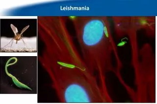
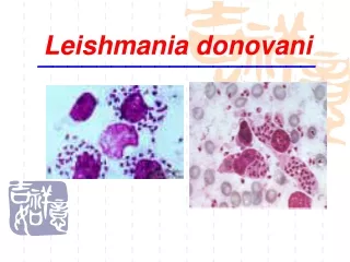
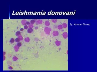
Leishmania donovani • Leishmania donovani causes visceral leishmaniasis. • It is also known as Dum-Dum fever,asian fever,Assam fever, • or infantile Splenomegaly in various part of the world. • Leishmania & donovan report the parasite simultaneously • in year 1903. • The sand fly,phlebotomusargentipes was identified as a • vector of disease by Indian Kala-azar commission.
Habitat • L.donovani is an obligate intracellular parasite • of man & other mammalian host. • Always found as intracellular amastigotes in • Reticulo-endothelial cells of the spleen • Bone marrow,liver, intestinal mucosa & mesenteric • lymph node.
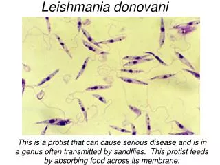
Morphology The parasite exist in two forms. Amastigote • Amastigote are found in man other mammalian host. • Found inside monocytes,polymarphonuclear leucocytes • or endothelial cells. • Also known as LD bodies, stained with giemsa or wright. • In gisemsa staining cytoplasm appear pale blue, • nucleus stained red
It is slender in rod shape & stained deep red. • Axoneme arises from the kinetoplast & extends • to margin of the body. • Vacuole, which is a clear unstained space, lies • alongside the axoneme. Promastigotes • It found in digestive tract of sand fly & in the • culture media. • The fully developed Promastigotes are long,slender & • spindle in shape.
Size: 15-25 micro meter in length & 1.5-3.5 in breadth. A single nucleus situated at center. Kinetoplast lies transversely near the anterior end. Flagellum is single delicate 15-28 micrometer in size. Leishman stain : cytoplasm appear in blue & nucleus in pink Kinetoplast bright red.
Culture& laboratory animals Biphasic media: Biphasic media to be developed for the culture. It consist of two part of salt agar & one part of defibrinated rabbit blood Liquid media: Schneider's’, grace’s & mituhasi-maramorosch media. This media do not contain blood.
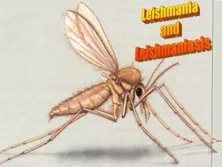
Laboratory animal: Chinese hamster & golden hamster are highly susceptible to Infection. Life cycle: Completes its life cycle in two different hosts 1.man & other mammals 2. Sand fly of genus Phlebotomus & Lutzomyia.
LIFE CYCLE OF LEISHMANIA DONOVANI Leishmania donovani in human blood Infection of sand fly via blood meal Multiplication by binary fission Migrating to salivary gland Infection of man by sand fly bite Reticulo - endothelial cycle Multiply by binary fission Enter blood circle
pathology Spleen: Macroscopic Grossly enlarge ,capsule thickened,soft in consistency. Microscopic:vascular spaces widely dilated. Liver: Macroscopic Liver is enlarged & congested. Microscopic: kupffer cell hyperplasia & cells are loaded with LD bodis
Bone marrow Increase number of plasma cell & LD bodies are in plenty. Intestine: ulcers Miscellaneous cloudy swelling of kidney may lead to albuminuria. Mode of transmission Bite of anthrophilic phlebotomus species. Man is the only reservoir of infection in Indian Kala-azar.
TRANSMITTED BY 1. bite of vector 2.blood transfusion 3. Congenital infection 4. Accidental inoculation in lab workers 5. Sexual intercourse. Diagnosis Specimen: spleen, bone marrow, liver, blood smear.
Method of examination Microscopy Culture Animal inoculation. Microscopy Stained by leishman , giemsa,or wright stains. • Culture • Spleen & bone marrow aspiration & buffy coates • of the blood show Promastigotes in culture. • NNN, Schneider drosophila medium for demonstration • of Promastigotes. • Incubate at 22-26 C for 1- 4 weeks.
Animal inoculation Inoculation of chinese & golden hamster by clinical Specimen may reveal parasite. • Serological test • CFT • IMMUNO- FLOURESCENT • ELISA. Molecular diagnosis DNA probes & PCR
Treatment • Pentavalent antimonials are the drug of choice. • Meglumine antimonate , sodium stibogluconate solution. • Prevention & control • Reducing of sand fly population using pesticides. • Reducing the infected dogs.