Understanding Spirochetes and Leptospirosis: Characteristics, Transmission, and Control
Learn about spirochetes, focusing on Leptospira and Leptospirosis. Explore their morphology, classification, epidemiology, pathogenicity, clinical findings, laboratory diagnosis, and control measures. Understand Treponema pallidum and Syphilis. Discover transmission methods, diseases, and prevention strategies.


Understanding Spirochetes and Leptospirosis: Characteristics, Transmission, and Control
E N D
Presentation Transcript
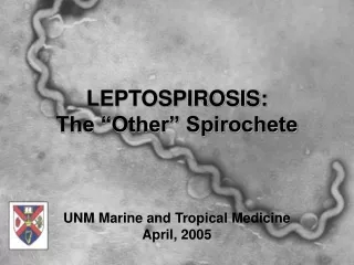
Inroduction • The spirochetes are a large heterogenous group of bacteria. • In some respects, this microorganism is similar to bacteria and protozoa. • They have cell wall and propagate by binary fission and move vigorously by the rotation and twisting of the endoflagella. They are sensitive to antibiotic.
Structural characteristics: ◇They are long, slender and spiral or helical-shaped, Gram-negative bacilli.
Protoplasmic cylinder Endoflagella Outer envelope • Structural characteristics: ◇There are endoflagella located between outer and inner membrane (envelope) and running parallel to the microbic body.
There are three genera whose members are human pathogens: • Leptospira • Treponema • Borrelia
Morphology • Leptospira are tightly coiled and flexible (5-15 µm long and 0.1-0.2 µmwide). One or two ends are usually bent to form hook-like shape. • While stained with silver stain (Fontana stain), they show a deep brown color.
Morphology • They are actively mobile, which is best seen using a darkfield microscope.
Classification • two species: • L. interrogans • causes human or animal diseases called leptospirosis • According to the antigenic differences of LPS and superficial glycoproteins, L. interrogans can be divided into at least 24 serogroups and 200 serotypes • L. biflaxa • Saprophytic, usually exists in water in nature.
Culture • Korthof medium with 10% rabbit serum • The optimal incubating temperature is about 28℃ • L. interrogans grows slowly • Sensitive to heating and various chemical agents • Can survive several months in wet soil and water • ◇L. interrogans has two chromosomes (one is large and the other is small).
Transmission • L. interrogans propagates in the kidney of infected patients or animals and can be shed in the urine. • Animal urine from infected rodents, infected farm animals (usually inapparent infection) • Contaminate soil and water • Enters body through healthy or broken skin. • Human leptospirosis. L. interrogans rapidly enters bloodstream to cause leptospiremia
Epidemiology • A common zoonosis: • A disease of animals that can be transmitted to humans. • Extensive animal hosts: • rats, mice, other wild rodents, swine, cattle, dog, sheep etc.
中华人民共和国地图 黑龙江省 吉林省 新疆维吾尔自治区 内蒙古自治区 辽宁省 甘肃省 北京市 % 天津市 E 山西省 河北省 % 宁夏回族自治区 青海省 % 山东省 % 西藏自治区 % 河南省 $ 江苏省 E % E 陕西省 % 安徽省 E % 四川省 湖北省 E E E % 浙江省 $ % E % 江西省 $ $ $ 湖南省 E % 贵州省 % % E 福建省 $ $ E E % $ 云南省 % 台湾省 广西壮族自治区 $ % E E 广东省 $ % $ E $ E % $ % % $ % $ % $ 海南省 E % $paddy planting area $ Leptospirosis area
Pathogenicity • Virulent factors produced by L. interrogans • LPS: lower toxicity than bacterial LPS • Hemolysin: unknown pathogenesis • Cytotoxicity factor (CTF) and cytopathic effect (CPE) substance: only show cytotoxicity • Pathological damage to the capillary endothelium is the main cause for disease.
Clinical finding • Leptospirosisaffects many internal organs e.g., lung, kidney, liver • The pathogenicity of different leptospiral serogroups is distinct. The clinical symptoms are quite different, from mild influenza-like clinical signs to death which is usually caused by pulmonary diffuse hemorrhage (PDH) • Macrophage can phagocytose L. interrogans, neutrophils can not
Laboratory Diagnosis • Peripheral blood in the first week of disease and urine from the second week on are collected as samples for detecting leptospire • Common diagnostic methods in clinic include direct darkfield microscopy and serological examination
Control • Penicillin is the first choice drug to treat leptospirosis • Multi-valent vaccine composed of whole dead cells of several leptospiral serovars is available. However side effect of the vaccine is relatively serious
Morphology of Treponema pallidum • Moves with endoflagella • There are 16 to 18 bends • Shows a deep brown color by silver stain. • Can be cultured in rabbit testicle but can not grow in vitro • Sensitive to temperature and dryness
Treponema pallidum in testis of infected rabbit
Transmission • via sexual contact • via placenta or during birth • via blood transfusion • Disease • Acquired syphilis • Congenital syphilis
I. Acquired Syphilis • Syphilis is the a most common STD in our country • This disease appears chronic and slowly progressive • Syphilis undergoes 3 stages
Stage 1: Primary Syphilis • Chancre appears (an area of ulceration/inflammation) usually in genital areas • The patient has influenza-like symptoms • The local lesion will heal within 4-6 weeks but the microbe spread systemically
Stage 2: Secondary Syphilis • Pale red rash (syphilid)appears on thepalms or soles or over theentire body • The sores around the genitals or anus secrete extremely infectious fluids • This stage will last 3-6 months
Syphilid Abdominal part palms soles
Stage 3: Latency Stage • No obvious symptoms and usually occurs 2-10 years after initial infection and last 3-6 months. • The infectious ability is decreased. • Later the complications in the skin, bones, central nervous system, heart and blood vessels appear, which frequently cause death. • The basic pathological lesion is chronic granuloma (Gumma).
Gumma (syphiloma) lesion in the heart Gumma (syphiloma) lesion in the skin
II. Congenital Syphilis • The disease can cause fetal death. • In infants, multi-organ deformity or latent infection are present.
I. Early Congenital Syphilis Runny nose Vesicular rash Osteochondritis
II. Late congenital Syphilis Saw-teeth Sabre shins Frontal bossing and saddle nose
Laboratory diagnosis • Samples • the secretions from chancre in Stage I and from syphilid in Stage II • the patient’s serum
Laboratory diagnosis Darkfield microscopy and fluorescence microscopy RPR (rapid plasma reagin) • Use cattle cardiolipin as the antigen to detect reagin (syphilis non-specific antibodies) in the serum TPPA (T. pallidum particle agglutination assay) or TPHA (T. pallidum hemagglutination assay) • micro-hemoagglutination assays for the detection of antibodies to Treponema pallidum
Treatment & Prevention • Penicillin • Tetracycline Erythromycin • No vaccine
Lyme disease is an emerging zoonosis mainly caused by Borrelia burgdorferi. In 1975, the disease was firstly found in a town named Lyme in the U.S.A. • Human is infected by bite of tickscarryingBorrelia burgdorferi. • The clinical characteristic is erythema chronicum migrans (ECM) at the bite site. • Borrelia burgdorferi can invade host cells, resist phagocytosis and produce endotoxin-like substance. Pathogenesis is considered to be closely associated with immunopathological injury because rarely the pathogen can be found in vivo.
Borrelia burgdorferiis a spiral microorganism. The microbe has variable numbers (7-11) of endoflagella to move actively.
Inn tick A kind of tick to transmit Borrelia burgdorferito human
Lyme Disease erythematous rash Erythema chronicum migrans (ECM)
Diagnosis & Treatment • Lyme disease is diagnosed based on clinical symptoms (e.g., ECM) and the possibility of exposure to infected ticks. • PCR and serological assays are helpful for clinical diagnosis. • Most of Lyme disease cases can be treated successfully with a few weeks of antibiotics (penicillin or tetracycline).