Download
1 / 52
830 likes | 1.71k Vues
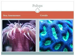
Colorectal polyps and polyposis syndromes. Division of Colorectal Surgery The Chinese University of Hong Kong. Intensive Surgery Course for Medical Year 5 (2006/2007). Colorectal polyps. visible protrusion above the surface of the surrounding normal large bowel mucosa

E N D
Colorectal polyps and polyposis syndromes Division of Colorectal Surgery The Chinese University of Hong Kong Intensive Surgery Course for Medical Year 5 (2006/2007)
Colorectal polyps • visible protrusion above the surface of the surrounding normal large bowel mucosa • Detected by endoscopy or by DCBE
Hyperplastic polyps • Majority of non-neoplastic polyps • Prevalence rates of 20-34% (autopsy and screening colonoscopy studies) • Predominantly located in the distal colon and rectum • Generally small (<0.5cm) in size
Adenomas – facts and figures • 70% of all colorectal polyps • Increase with age (33% of population by 50yr, and in 50% by 70yr) • 70% located in the left colon • 70% are solitary (30% synchronous) • 70% are small (<1cm in size) • 7% have severe dysplasia, 3-5% have invasive cancer
Adenoma CRC Adenoma-carcinoma sequence 10 years Regardless of aetiology, most CRC arise from adenomas
Factors determining risk of malignant transformation within adenomas
Percent of adenomas containing invasive cancer by size and histology
Malignant colorectal polyp • Polyp that contains invasive cancer • Malignant cells that have invaded through the mucularismucosa into the submucosa mm
Management of colorectal polyps (1)Factors Location: colon or rectum Morphology: pedunculated or sessile Histology: benign or malignant
Management of colorectal polyps (2)Excision Pedunculated Colonoscopic polypectomy usually possible Sessile Colonoscopic polypectomy if possible (larger polyps may require piecemeal removal) Endoscopic removable not possible operative removal • Colon: colectomy • Rectum: staged with EUS or MRI • Benign / Early malignant (T1No) : Transanal local excision or TEMS (may need further radical surgery) • Other malignant : radical excision (APR /anterior resection)
Benign Surveillance colonoscopy Malignant Depends on histological characteristics Management of colorectal polyps (3)Definitive Mx (histology) Radical Surgery
Surveillance after polypectomyBenign polyps Guidelines for colonoscopy surveillance after polypectomy: a consensus update by the US Multi-Society Task Force on Colorectal Cancer and the American Cancer Society (2006)
Malignant PolypFactors determining need of radical surgery Histology • Poorly differentiated • Margin <2mm • Stalk invasion • Lymphovascular invasion Increase risk of recurrence and LN 2o
Familial adenomatous polyposis (FAP) • 1% of all CRC • Present in about 1 in 8000 births • Autosomal dominant with near 100% penetrance
FAP • >100 adenomas • Patients develop adenomas by the mean age of 16 years, and CRC by 39 years • Adenomas form early, but it takes 20-30 years to develop CRC from adenomas • Disease of abnormal tumour initiation
Molecular genetics of FAP • Caused by mutations of APC gene (tumour suppressor gene) on chromosome 5q21 • Encodes for a protein, which functions in cell adhesion and signal transduction • Mutations will result in truncated protein and affect cell growth
Genotype vs. phenotype Affected part of gene Clinical Presentation Extracolonic manifestations Cell adhesion and structural molecules
CHRPE Extracolonic manifestations • Congenital hypertrophy of retinal pigmented epithelium (CHRPE) • Osteomas, desmoid tumours, epidermoid cysts (Gardner’s syndrome) • CNS malignancies including medulloblastoma and glioblastoma (Turcot’s syndrome) • Duodenal, hepatobiliary-pancreatic, thyroid tumours
Gardner’s syndrome Desmoid Chest fibroma Mandibular osteoma Skull osteoma
Attenuated FAP (AFAP) • Variant of FAP • <100 adenomas • Late age-of-onset (adenomas at 44; CRC at 56) • Proximal distribution of adenomas *Colonoscopy for surveillance *Infrequent involvement of the rectum supports the role of total colectomy and IRA
Mutation Protein truncation test DNA sequencing Diagnosis of FAP Endoscopy Genetic tests
Screening of FAP • Genetic screening of family members for APC mutations • Annualflexible sigmoidoscopy beginning at age 10-12 until age 40, then every 3-5 years *If polyposis is present, colectomy should be considered • OGD every 1-3 years is also recommended to evaluate for upper GI adenomas
CRC FAP Prophylactic colectomy for FAP • The aim of surgical treatment of FAP is to intervene in the adenoma-carcinoma sequence by removing the adenomas before the transformation to malignancy occurs
Standard surgical treatment Restorative proctocolectomy with ileal pouch-anal anastomosis Suitable for most patients with FAP
Other surgical options Total colectomy with ileorectal anastomosis (IRA) Proctocolectomy with ileostomy low rectal cancers poor sphincters Desmoid tumors Attenuated FAP
Medical treatment of FAP? • Sulindac (NSAID) and celecoxib (COX-2 inhibitor) shown to control and reduce the number of colorectal adenomas in FAP • Not definitive treatment • Temporizing treatment (eg when surgery needs to be delayed) • May control pouch and rectal polyposis after initial prophylactic surgery
Hereditary nonpolyposis colorectal cancer (HNPCC) Dr. Henry Lynch first described the term ‘cancer family syndrome’ in 1966 (later renamed as Lynch syndrome and HNPCC) Dr. A. S. Warthin and the first HNPCC pedigree, ‘the family G’ 1895
HNPCC • 2-5% of all CRC • Autosomal dominant • 70-80% penetrance • It takes only 3-5 years to develop CRC from adenomas Accelerated progression
HNPCC related extracolonic tumors Endometrial cancer is the most common extracolonic malignancy
Diagnosis: Amsterdam criteria 1 Due to lack of phenotypic markers like polyps Diagnosis is based on family history of CRC only • One member less than 50 years of age • Two involved generations • Three family members affected, one of whom is a first-degree relative of the other two
Diagnosis: Amsterdam criteria 2 Same as Amsterdam 1 but includes all HNPCC related tumors
Molecular genetics of HNPCC HNPCC is caused by mutations of DNA mismatch repair (MMR) genes Survey DNA for replication errors
Molecular genetics of HNPCC • Mutations of these MMR genes will result in replication errors during DNA synthesis (microsatellite instability) leading to acceleration of genetic mutations • HNPCC patients develop adenomas at the same rate as the general population • Once these adenomas develop, however, defective DNA repair ensues and mismatches accumulates • Thus, it takes only 3-5 years to develop CRC from adenomas
Demonstration of MSI DNA Normal Tissue DNA Tumor Tissue Microsatellite Markers Amplify by Polymerase Chain Reaction Compare normal and tumor profiles of amplified microsatellite on gel to detect genetic mutations in these microsatellites
MSS Tumor BAT 25 D17S250 BAT 26 D2S123 D5S346 NORMAL MUCOSA TUMOR
MSI Tumor NORMAL TUMOR
Microsatellite instability testing Negative: Stop? Positive Immunohistochemistry Normal Stop? MLH1 MSH2 Sequence MLH1 No protein Sequence MSH2 No protein HNPCC: Mutation detection for MLH1 and MSH2
Screening of HNPCC • Colonoscopy every 2 years starting at ages 20-25 or 5 years younger than the earliest diagnosis of CRC whichever is earlier until 40yr , and then annually • Flexible sigmoidoscopy is notacceptable, due to the proximal location of tumours • Transvaginal US and endometrial aspiration annually starting at ages 25-35 years are also recommended
Surgical treatment of HNPCC • Total colectomy with ileorectal anastomosis • Restorative proctocolectomy with ileal pouch-anal anastomosis • Segmental colectomy notrecommended because of high rate of metachronous CRC • TAHBSO for endometrial cancer
Peutz-Jeghers syndrome • Incidence: 1 in 200,000 persons Autosomal dominant • Mutations of the STK11 gene on chromosome 19 • Characterized by perioral pigmentationsand hamartomatous polyps throughout the GI tract • GI and non-GI cancers are common