Download
1 / 34
440 likes | 2.2k Vues
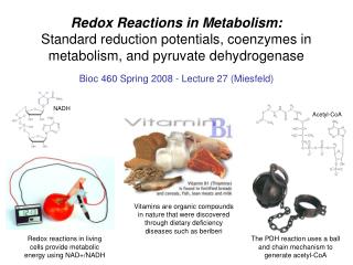
Redox Reactions in Metabolism: Standard reduction potentials, coenzymes in metabolism, and pyruvate dehydrogenase. Bioc 460 Spring 2008 - Lecture 27 (Miesfeld). NADH. Acetyl-CoA. Vitamins are organic compounds in nature that were discovered through dietary deficiency diseases such as beriberi.

E N D
Redox Reactions in Metabolism:Standard reduction potentials, coenzymes in metabolism, and pyruvate dehydrogenase Bioc 460 Spring 2008 - Lecture 27 (Miesfeld) NADH Acetyl-CoA Vitamins are organic compounds in nature that were discovered through dietary deficiency diseases such as beriberi Redox reactions in living cells provide metabolic energy using NAD+/NADH The PDH reaction uses a ball and chain mechanism to generate acetyl-CoA
Key Concepts in Redox Metabolism 1. Reduction potentials are a measurement of electron affinity. Compounds with a very high affinity for electrons are oxidants, e.g., O2, and have a positive reduction potential (Eº’>0). Very strong reductants are compounds that readily give up electrons, e.g., NADH, and have a negative reduction potential (Eº’<0). Electrons flow from reductants to oxidants (electrons flow toward compounds with higher Eº’ values).
Key Concepts in Redox Metabolism 2. Coenzymes are organic compounds that provide reactive chemical groups to enzymes. Many coenzymes were discovered as vitamins through the study of dietary deficiency diseases. Most coenzymes, such as nicotinamide adenine dinucleotide (NADH) and thiamin pyrophosphate (TPP), are noncovalently associated with enzymes.
Key Concepts in Redox Metabolism 3. The pyruvate dehydrogenase (PDH) complex is a mitochondrial metabolic machine that converts pyruvate to acetyl-CoA in a favorable reaction (Gº’ = -33.4 kJ/mol). The PDH reaction is in the mitochondrial matrix and captures decarboxylation energy in the form of NADH.
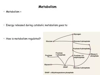
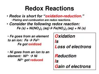
Redox reactions transfer electrons Redox reactions (oxidation-reduction) in the citrate cycle are a form of energy conversion involving the transfer of electron pairs from organic substrates to the carrier molecules NAD+ and FAD. The energy available from redox reactions is due to differences in the electron affinity of two compounds and is an inherent property of each molecule based on molecular structure. Coupled redox reactions consist of two half reactions: 1) an oxidation reaction (loss of electrons) 2) a reduction reaction (gain of electrons).
Conjugate redox pairs Compounds that accept electrons are called oxidants and are reduced in the reaction, whereas compounds that donate electrons are called reductants and are oxidized by loss of electrons. Each half reaction consists of a conjugate redox pair represented by a molecule with and without an electron (e-). Fe2+/Fe3+ is a conjugate redox pair in which the ferrous ion (Fe2+) is the reductant that loses an e- during oxidation to generate a ferric ion (Fe3+) the oxidant: Fe2+ <--> Fe3+ + e- Similarly, the reductant cuprous ion (Cu+) can be oxidized to form the oxidant cupric ion (Cu2+) plus an e- in the reaction: Cu+ <--> Cu2+ + e-
Conjugate redox pairs Two half reactions are combined to form a redox reaction. For example, the transfer of an e- from from Fe2+ (the reductant) to Cu2+ (the oxidant) to form Fe3+ and Cu+. Fe2+ <--> Fe3+ + e- Cu2+ + e- <--> Cu+ Fe2+ + Cu2+ <--> Fe3+ + Cu+ The Fe was oxidized and the Cu was reduced in a redox reaction in which the e- was the shared intermediate. This Fe-Cu redox reaction takes place within the cytochrome c oxidase complex in the electron transport system of the inner mitochondrial membrane.
Aerobic respiration is the transfer of electrons from glucose to O2 to form CO2 and H2O The more electrons a carbon atom has available to donate, the more reduced (less oxidized) it is. Hydrogen is less electronegative than carbon, and therefore electrons in C-H bonds are considered "owned" by the carbon. Oxygen is more electronegative than carbon and the electrons in C-O and C=O bonds are all "owned" by the oxygen atom.
Redox reactions in the citrate cycle involve the transfer of e- pairs to generate NADH and FADH2 The reduction of NAD+ to NADH involves the transfer of a hydride ion (:H-), which contains 2 e- and 1 H+, and the release of a proton (H+) into solution NAD+ + 2 e- + 2 H+ <--> NADH + H+ In contrast, FAD is reduced by sequential addition of one hydrogen (1 e- and 1 H+) at a time to give the fully reduced FADH2 product FAD + 1 e- + 1 H+ <--> FADH + 1 e- + H+ <--> FADH2 Enzymes that catalyze biochemical redox reactions are strictly called oxidoreductases, however, since most oxidation reactions involve the loss of one or more hydrogen atoms, they are often called dehydrogenases.
Reduction potential (E) is a measure of the electron affinity of a given redox pair Biochemical standard reduction potentials (Eº’) are determined under standard conditions using an electrochemical cell that measures the relative e- affinity of a test redox pair compared to the hydrogen half reaction. Two half cells are connected by a galvanometer which measures the flow of electrons between two electrochemical cells. An agar bridge between the two half cells allows ions to flow and balance the charge to keep the electron circuit intact. Fe3+ has a higher e- affinity than H+
Standard reduction potentials are expressed as half reactions and written in the direction of a reduction reaction. Redox pairs with a positive Eº’ have a higher affinity for electrons than redox pairs with a negative Eº’. Electrons move from the redox pair with the lower Eº’ (more negative) to the redox pair with the higher Eº’ (more positive). The hydrogen half reaction is set as the standard with a Eº’ = 0 Volts.
The amount of energy available from a coupled redox reaction is defined as Eº’ By convention, the Eº' of a coupled redox reaction is determined by subtracting the Eº' of the oxidant (e- acceptor) from the Eº' of the reductant (e- donor) using the following equation: Eº' = (Eº'e- acceptor) - (Eº'e- donor) The Eº' for a coupled redox reaction is proportional to the change in free energy Gº' as described by the equation (n is the number of e-): Gº' = -nFEº' If Eº' > 0, then the reaction is favorable since Gº' will be negative. A coupled redox reaction is favorable when the reduction potential of the e- acceptor is more positive than that of the e- donor.
Calculating the Gº’ for a citrate cycle oxidation reaction using the Eº’ of the half reactions • The oxidative decarboxylation of isocitrate by the enzyme isocitrate dehydrogenase in the third reaction of the citrate cycle: • Isocitrate + NAD+ <--> -ketoglutarate + CO2 + NADH + H+ • Using the Eº’ values from the table with the half reactions as reductions: • NAD+ + H+ + 2 e- ---> NADH (Eº’ = -0.32 V) -ketoglutarate + CO2 + 2 e- + 2 H+ ---> isocitrate (Eº’ = -0.38 V) • And now calculate Eº’ considering that NAD+ is the e- acceptor and isocitrate is the e- donor (electrons move from low Eº’ to higher Eº’): • Eº' = (Eº'e- acceptor) - (Eº'e- donor) • Eº' = (-0.32 V) - (-0.38 V) =+0.06 V
Another way to get the same answer • If it makes more sense to you to write the two half reactions in the direction of the overall net reaction, then simply reverse the Eº’ value for the isocitrate oxidation and add the two Eº’ values together: • Writing each half reaction in the direction of the net reaction: • NAD+ + H+ + 2 e- ---> NADH (Eº’ = -0.32 V) isocitrate ---> -ketoglutarate + CO2 + 2 e- + 2 H+(Eº’ = +0.38 V) • Isocitrate + NAD+ <--> -ketoglutarate + CO2 + NADH + H+ • Eº' = (-0.32 V) + (+0.38 V) =+0.06 V This is the method used in the Berg textbook (pg. 508), although in that case, they calculate the Gº’ values first, and then add the Gº’ values together.
Now we can use this Eº’ value to calculate the Gº’ for the reaction • Gº' = -nFEº' • Gº' = (-2) •(96.48 kJ/molV) • (+0.06 V) • Gº' = -11.6 kJ/mol • A value for Gº’ < 0 confirms that this coupled redox reaction is favorable, i.e., it is favorable to oxidize isocitrate and reduce NAD+. • In order to calculate the actual reduction potentials for conjugate redox pairs, you need to use the Nernst equation and know the actual concentration of the oxidant (e- acceptor) and the reductant (e- donor) inside the cell (the mitochondrial matrix in this case): E = Eº' + RT ln [e- acceptor] nF [e- donor]
Pyruvate destined for the citrate cycle, or fatty acid synthesis, is converted to acetyl CoA by pyruvate dehydrogenase (PDH). Acetyl-CoA has only two metabolic fates in the cell, and therefore, its production by PDH must be tightly regulated. • acetyl-CoA can be metabolized by the citrate cycle to convert redox energy to ATP by oxidative phosphorylation • acetyl-CoA can be used as a form of stored energy by conversion to fatty acids that are transported to adipocytes (fat cells) as triglycerides.
The pyruvate dehydrogenase complex catalyzes the oxidative decarboxylation of pyruvate to form CO2 and acetyl-CoA in a reaction that requires three enzymes (E1, E2, and E3), and five coenzymes (NAD+, FAD, CoA, TPP, and lipoic acid), that work together to catalyze five linked redox reactions. • Pyruvate + CoA + NAD+ ---> acetyl-CoA + CO2 + NADH • Gº’ = -33.4 kJ/mol
Coenzymes provide additional chemical groups to enzymes that facilitate catalysis Why are human vitamin deficiencies rare in developed countries?
Nicotinamide adenine dinucleotide (NAD+) NAD is derived from the water-soluble vitamin niacin which is also called vitamin B3. NAD+, and its phosphorylated form NADP+, are involved in over 200 redox reactions in the cell which are characterized by the transfer of 2 e- as hydride ions (:H-). Catabolic redox reactions primarily use the conjugate redox pair NAD+/NADH and anabolic redox reactions use NADP+/NADPH. Note that the "+" charge does not refer to the overall charge of the NAD molecule, but rather only to the charge on the ring N in the oxidized state.
Nicotinamide adenine dinucleotide (NAD+) Severe niacin deficiency causes the disease pellagra which is associated with diets consisting primarily of cultivated corn. Pellagra is rare in Mexico because corn used for tortillas is traditionally soaked in lime solution prior to cooking and this releases niacin from its bound form upon heating. Since corn obviously contains niacin, why would eating corn "cause" pellagra?
Flavin adenine dinucleotide (FAD) FAD is derived from the water-soluble vitamin riboflavin which is also called vitamin B2. Riboflavin is destroyed by light, therefore, milk is now stored in light-tight containers. FAD is reduced to FADH2 by the transfer of two electrons in the form of hydrogen atoms. FAD can accept one electron through a reduced intermediate, semiquinone (FADH).
acetate group Coenzyme A (CoA) CoA is derived from the water-soluble vitamin pantothenic acid which is also called vitamin B5. CoA is absolutely essential for life as it is required for energy conversion by the citrate cycle, it is also a cofactor in fatty acid, acetylcholine, heme and cholesterol biosynthetic pathways. The primary role of CoA is to function as a carrier molecule for acetate units in the form of acetyl-CoA.
Coenzyme A (CoA) Acetyl-CoA consists of a central pantothenic acid unit that is linked to a functional -mercaptoethylamine group. Acetate is covalently attached to CoA through an activated thioester bond which has a high standard free energy of hydrolysis. Attachment of acetate units to the reduced form of CoA requires reactions with high Gº' values, for example, PDH (Gº' = -33.4 kJ/mol).
Thiamin pyrophosphate (TPP) FAD is derived from the water-soluble vitamin thiamin (or thiamine) which is also called vitamin B1. A carbon atom on the thiazole ring of TPP is the functional component of the coenzyme and is involved in aldehyde transfer. Thiamin is phosphorylated by the enzyme thiamin kinase in the presence of ATP to form thiamin pyrophosphate (TPP) and AMP.
Fish and silkworms contain the enzyme thiaminase, which degrades thiamin. Cooking these foods destroys the thiaminase. Thiamin pyrophosphate (TPP) Thiamin deficiency is the cause of beriberi and has been found in populations that rely on white polished rice as a primary source of nutrition. Milling rice removes the bran which contains thiamin.
High levels of -lipoic acid are found in broccoli, spinach, and tomato. -Lipoic acid (lipoamide in proteins) The role of -lipoic acid in metabolic reactions is to provide a reactive disulfide that can participate in redox reactions within the enzyme active site. -Lipoic acid is not considered a vitamin because it is synthesized at measurable levels in humans.
-Lipoic acid (lipoamide in proteins) Lipoamide, the naturally occurring form of -lipoic acid, is a covalent linkage of -lipoic acid to a lysine -amino group on proteins. The long hydrocarbon chain bridging -lipoic acid and lysine provides a flexible extension to the reactive thiol group. The E2 subunit of the pyruvate dehydrogenase complex contains the lipoamide at the end of a polypeptide tether which functions as a "ball and chain" that moves the lipoamide back and forth across a 50 Å span in the interior of the complex.
The pyruvate dehydrogenase (PDH) complex is a highly efficient metabolic machine The conversion of pyruvate to acetyl-CoA by the pyruvate dehydrogenase complex is an oxidative decarboxylation reaction that represents another amazing example of protein structure and function. The eukaryotic pyruvate dehydrogenase complex contains multiple subunits of three different catalytic enzymes that work together as a metabolic machine. Note that TPP, lipoamide, and FAD are all regenerated.
The pyruvate dehydrogenase (PDH) complex is a highly efficient metabolic machine Three of the coenzymes are covalently linked to enzyme subunits, with TPP attached to the E1 pyruvate dehydrogenase subunit, lipoamide is the functional component of the E2 dihydrolipoyl transacetylase subunit, and FAD is covalently bound to the E3 dihydrolipoyl dehydrogenase subunit. The two other coenzymes, CoA and NAD+, are transiently associated with the E2 and E3 complexes, respectively.
The pyruvate dehydrogenase (PDH) complex is a highly efficient metabolic machine The pyruvate dehydrogenase reaction can be broken down into five distinct catalytic steps: Decarboxylation Transfer of the acetyl group to lipoamide Formation of acetyl-CoA Redox reaction to form FADH2 Redox reaction to form NADH 5 4 1 2 3
The pyruvate dehydrogenase (PDH) complex is a highly efficient metabolic machine The E1, E2 and E3 subunits of the mammalian PDH complex are packed together in a huge ~400 Å diameter sphere with a combined molecular weight of ~7800 kDa. The stoichiometry of the E1:E2:3 subunits (22:60:6) is consistent with there being 60 active sites in the pyruvate dehydrogenase complex.
The pyruvate dehydrogenase (PDH) complex is a highly efficient metabolic machine The lipoamide moiety of the E2 subunit is attached near the end of a ~200 amino acid long segment of the protein that functions as both a structural linker connecting the E2 and E1 subunits, and as a type of lipoamide "ball and chain."
Arsenite is a naturally occurring inhibitor of lipoamide Inadvertent ingestion arsenite can lead to an untimely death by irreversibly blocking the catalytic activity of lipoamide-containing enzymes such as the PDH and -ketoglutarate dehydrogenase complexes. Chronic arsenic poisoning can come from environmental sources such as arsenic-contaminated drinking water and result in the appearance of ulcerous skin lesions and an increased risk of a variety of cancers.
Arsenite is a naturally occurring inhibitor of lipoamide Since the 1990s it has been documented that millions of people in India have been chronically exposed to toxic levels of arsenic in contaminated drinking water obtained from shallow hand-pumped wells. During the 1970s and 1980s, UNICEF and other relief organizations helped drill thousands of wells in small Indian villages as an humanitarian effort to circumvent public water supplies that had become biologically contaminated. About ten years later when large numbers of villagers in the Ganges delta region developed skin lesions and cancers, it was realized that these wells contained water with toxic levels of arsenic. Massive efforts were undertaken to close down contaminated wells and to develop purification systems to reduce the arsenic to safe levels in other water supplies.