Download
1 / 44
440 likes | 692 Vues
The HPV-16 E7 Oncogene Sensitizes Malignant Cells to IFN-alpha-Induced Apoptosis.

E N D
The HPV-16 E7 Oncogene Sensitizes Malignant Cells to IFN-alpha-Induced Apoptosis Thyrell, L., O. Sangfelt, B. Zhivotovsky, K. Pokrovskaja, Y. Wang, S. Einhorn, D. Grander.2005. The HPV-16 E7 Oncogene Sensitizes Malignant Cells to IFN-alpha-Induced Apoptosis. Journal of Interferon and Cytokine Research 25:63-72.
HPV-What is it?Human Papillomavirus • Figure A: Double stranded HPV viral strain.
Human Papillomavirus (HPV), is the member of a family of viruses that cause abnormal tissue growth and other changes to cells HPV can be categorized into two subgroups, according to how they infect the genital tract A) “Low Risk” or B) “High Risk” HPV and its Categorization
“What does Low Risk HPV do” • (Figure C:“low risk” HPV- genital warts on ano-vaginal area). (http://www.lib.uiowa.edu/hardin/md/genitalwarts.html 2005).
Does “High Risk” HPV cause Cervical Cancer?YES!!!! Figure D: Normal cervix Figure E: “High risk” HPV that causes cervical cancer
Viral Life CycleLytic or Lysogenic Viral Life Cycle? • HPV has a Lysogenic Viral Life Cycle • 1). The virus attaches itself and injects its DNA into the cell. • 2). The viral DNA attaches itself to the host DNA, becoming a new set of cell genes called a prophage. • 3). When the host cell divides, this new gene is replicated and passed to new cells. This causes no harm to the cell, but may alter its traits.
Figure:B-Lysogenic viral cycle-Bacteria is representing viral cell
Is there an association between Lysogenic Viruses and Cervical Cancer? • All cells have genes which control their growth and differentiation. • Many of these genes are oncogenes because if they are expressed abnormally they cause uncontrolled growth and differentiation. • How does This Happen? • HPV viral genome activates proto-oncogenes, which can occur when certain viruses invade a normal cell. • Once the correct mutation has occurred to convert a proto-oncogene into a carcinogenic form (called an oncogene), cancer results. • Activation of proto-oncogenes to form oncogenes disrupts the normal cycle of the cell, and causes cancer (Zur Hausen 1996).
How Does HPV cause Cancer? • Normal Cell TRANSFORMSto Cancer Cell BY: • Activation of Oncogenes • The Inactivation of Tumor Suppressor Genes
The Role of Interferons • The Interferons are a family of proteins that have anti-viral functions. • IFN (Interferons) perform Cellular responses for anti viral function: • A). inhibition of proliferation (stopping the growth) • B). Immuno-regulatory effects (regulation the immune response), have been proposed as methods that the interferon use in the antiviral function
How does IFN inhibit proliferation?APOPTOSIS-Programmed Cell Death • Induction of apoptosis are induced by IFNs, and apoptotic induction by this protein, has been proposed to be of importance for both its anti-tumural in addition to its anti-viral responses • The IFN-inducing apoptosis function enables the protein to destroy the virus that is causing the tumor growth.
The Association between IFN-Alpha and the Caspase family • In a study by Thyrell et al, the aim of the study was to delineate the pathways activated during IFN-alpha induced apoptosis in malignant cell lines • Activation of the Caspase family is critical in order to complete Apoptosis • IFN-alpha + Caspase family =INDUCED APOPTOSIS
Cell Cycle • Figure :E-The cell cycle consisting of two overall phases, Division and Interphase.
Figure:F(www.sdsc.edu/ Jukebox/image7.html) p53 proteins have been found to suppress the growth of tumors and protect genes from mutating excessively. In this image, the p53 protein is shown on the left, and the helical DNA is shown on the right What is the Role of p53 in the cell cycle???
P53 tumor suppressor and its effects on the cell cycle • Figure:G-showing the interdependence of cyclin-kinase proteins, in order for cell cycle progression. • Cyclin + Kinase = Cell cycle progression • Cyclin + p53 = Stopping of cell cycle progression
Oncogenes E6/E7, activated by the HPV virus, Inhibit Apoptosis! Problem: • Oncoproteins in HPV activate an over-expression of Cyclin A and E. 2. Over-expression disrupts Tumor suppressor proteins p53 and Rb so that the proteins cannot respond to DNA damage and cannot halt the cell cycle in the G1 phase. As a result the damaged DNA continue to progress through the cell cycle
The effect Cyclins have on the Cell Cycle • Figure H: The malignant phenotype of the high risk HPV 16 depends on the viral oncoproteins E6 and E7, which alters the overexpression of the cyclins A and E, and cause a negative effect on the regulation of the cell cycle (Phelps et al 1992).
Phosphoylated pRb (no mutation cell) in normal cell cycle-release E2F protein to start DNA replication Dephosphorylated pRb (mutated cell) stops DNA replication by binding to E2F protein. Viral oncoprotein bind to dephosphorylated pRb form, which is also the form E2F protein binds to. Oncoprotein shoves away E2F protein E2 is released- causes cell cycle to continue (which induces transcription) Mutated cells continue to replicate, and are never again able to stop replicating because the oncoprotein stays bound to pRb How does pRb tumor suppressor cause proliferation
Let’s Review! • E7 has an ability to deregulate the control of cell cycle progression allowing cells to continue through G phase and enter S phase (replication) without inducing cell death to the mutated virus genes. • The oncogenic potential of the E6 and E7 proteins from the high-risk HPVs is due predominantly to the ability of these viral proteins to target and inhibit the activity of the cellular p53 and Rb tumor suppressor proteins respectively. • The ability of HPV to target both Rb and p53 allows the virus to stimulate cell replication, which also results in the replication of the viral genome while inhibiting apoptosis in the infected cell
E6 oncoprotein and its association with p53 • Cells expressing E6 lose the G1 checkpoint due to the ability of E6 to disrupt p53 pathway by degradation of p53 and are therefore resistant to p53-mediated apoptosis (Koromilas et al 2001). • The ability of E6 to mediate p53 degradation is very important considering that p53 places a zero tolerance on cellular abnormalities through mediating apoptosis and preventing cell proliferation. • Therefore in order for tumor cells or viral oncogene expressing cells to survive, the p53 tumor suppressor protein must be lost through gene mutation or viral protein interaction.
Why did Teaira ramble on about all this back ground information? • Because as stated before, the activation of proto-oncogenes to form oncogenes disrupts the normal cycle of the cell, and causes cancer. • The research performed by Thyrell et al, used the emerging research of the possibility that because key proteins are altered in malignant cells, the malignant genotype (HPV-16 E7) may be responsible in the cell’s sensitivity to IFN-alpha-induced apoptosis (Zur Hausen et al 2005).
Materials and Methods • In the experiment, Thyrell used the cell lines derived from mice, which contained the v-myc retrovirus-induced murine T cell lymphoma J3D6 • The test was to determine if the introduction of the E7 will increase sensitivity to the interferons’s ability to induce apoptosis. • The experimental group received transfected pbabe E7-puro vector ( it received the E7 gene) into the J3D6 cell line, called the J3D E7. • The control group was introduced to an empty vector containing no E7 puro vector, called the J3D puro.
Northern Blot Analysis • A northern blot is very similar to a southern blot except that it is RNA rather than DNA. The RNA is extracted, run on a gel and transferred to a filter membrane. There are three types of RNA: • 1. tRNA-Transfer RNA- which is responsible for the assembly of polypeptide chains • 2. rRNA-Ribosomal RNA-part of the structure of ribosomes • 3. mRNA-messengerRNA-the product is DNA transcription and used for translation of a gene into a protein. • *IT is mRNA which is isolated and hybridized in northern blots
In the experiment, the northern blotting analysis was able to measure the expression of HPV E7 by extracting total cellular RNA from the mock-transfected clone and the E7-transfected clones (Thyrell et al 2005) Figure:1
Figure one- once again the figure is simply showing the presence of expression E7 that was transfected to the mouse lymphoma cell lines (sub-clones 2-5), called J3D- E7 (experimental group) and the presence of the mouse lymphoma cell lines without the E7 expression (sub-clone 1), called J3D-puro (control group).
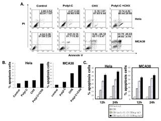
Flow Cytometry Analysis of DNAFigure:2 • Flow cytomety-was used to perform: • - DNA-fragmentation analysis • -Terminal deoxynucleotidyl transferase-mediated dUTP nick-end labeling (tunnel) analysis • -Analysis of Cellular DNA content, • -In viro caspase Assay • -flow cytometry analysis of Bak activation • Figure A: Flow Cytometry Software
Figure 2-The sub-clones J3D-E7 (experimental), along with sub-clone J3D-puro cells (control) were treated with the presence and absence of IFN-alpha for 72 hours
Let’s Review • J3D-E7 in the presence of IFN-alpha produced the most accumulation of sub-G1 DNA. • Increased DNA at sub G1 phase indicate induced apoptosis, because the cell cycle has stopped, enabling the expression of E7 to sensitize IFN-alpha to induce apoptosis
Expression of E7 sensitizes malignant cells to IFN-alpha apoptosis • The apoptotic morphology of the J3D-E7-transfected sub-clones were investigated by Histogram analysis by the observance of visible stained cells • DNA Histogram Analysis confirmed that the J3D-E7 sub-clones had the appearance of a large percentage of cells with typical apoptotic nuclear morphology, especially in all three J3D-E7 sub-clones (data was neither provided nor directed). • Significance-Stained nuclei = apparent morphological cells =Induced apoptosis
Annexin V Analysis • Figure:3
The experiment ultimately isolated and revealed the mechanisms associated with E7 as well as IFN-alpha. • The first detectionconfirmed the ability of the J3D-E7 cells to undergo apoptosis without the presence of IFN-alpha, signifying the function of the E7 expression to naturally sensitive malignant cells to apoptosis. • The Second revelation demonstrated IFN-alpha’s function in spontaneously inducing apoptosis when E7 expression is not available, signifying its ability to naturally induce apoptosis when confronted with malignant cells (Thyrell et at 2005).
DNA fragmentation correlates with the Induction of IFN-alpha-apoptosis
Sensitization to Apoptosis by E7 Correlates to Caspase Activation • IFN-alpha + Activation of Caspases = Induced Apoptosis • Caspase-8 was the vital initiator of the Caspase Cascade that subsequently activates the caspase family to carry out its duty in generating cell death. • The knowledge acquired pertaining to the association of the Caspase family and IFN-alpha-induced apoptosis, enabled Thyrell to investigate the involvement of Caspases in sensitization to apoptosis by the expression of E7.
Significance of Figure Five: • Once again, these results illustrate the lack of caspase activation in J3D-puro cells which means that there was a lack of INF-Alpha. • If there was a lack of IFN-alpha then there was a lack of apoptosis. • This makes sense, because there was a lack of expression E7 in puro cells (control), which does not sensitize IFN-alpha to malignant cells to induce apoptosis. • The increased activation of caspases in J3D-E7 transfecting cells, indicate the involvement of E7 oncoprotein has on the sensitization to IFN-alpha in inducing apoptosis due to the evidence that the presence of caspases is interdependent to IFN-alpha, for they are both tumor suppressors that need each other to carry out the completion of programmed cell death (Figure 5)(Thyrell et al 2005).
Expression of E7 in malignant cells Sensitizes to IFN-alpha induced Activation of the pro-apoptotic protein Bak • Recent studies indicate that IFN-alpha as well as DXR induces activation of the pro-apoptotic protein Bak prior to execution of apoptosis • Anthroacyclin Doxorubicin (DXR) is an antitumor antibiotic that interacts with DNA, affecting DNA and RNA synthesis • DXR induced apoptosis typically involves cytochrome c release from the mitochondria and subsequent caspase activation
Release of cytochrome c from the mitochondria intra-membrane space to the cytoplasm is commonly mediated by Bcl-2 family proteins Bak and Bax Therefore DXR activates pro-apoptoticmembers of the BCL-2 family. Thyrell used the innovative knowledge on DXR and associated DXR-induced apoptosis regulatory protein (BAK) to construct another test to understand the association with E7 and the sensitization to IFN-alpha induced apoptosis of J3D-E7 and J3D puro cells for 24 and 48 hours in the presence of IFN and DXR respectively DXR
HUH??? • IFN(Tumor suppressor) + DXR(Tumor suppressor) • = production of Bak • = induced apoptosis • E7 sensitizes IFN, which in return produced Bak, and completes induced apoptosis • DXR is proven to be a good tumor suppressor in malignant cells because it induces apoptosis, but with a patient who has high risk HPV with the expression of E7, the E7 will not sensitize DXR into apoptosis.
HUH?? • E7 has no effect on the sensitization of DXR, DXR is going to activate the same amount of caspase in both the control and experimental, in efforts to complete apoptosis
Conclusion • The scientific field has previously thought that the High risk HPV oncoprotein E7, disrupts all Tumor suppression genes, and only causes proliferation of abnormal cells. • Thyrell’s research on the expression of E7 and its sensitization on IFN-alpha to induce apoptosis, can now offer new treatments against HPV and associated cervical cancer. • In addition, other pathways, such as Caspase activation, has been recently looked into, in order to attempt to find another way to sensitize IFN-alpha to induced apoptosis on malignant cells