Download
1 / 34
340 likes | 556 Vues
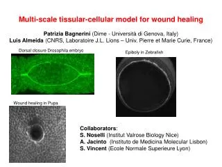
Multi- scale tissular -cellular model for wound healing Patrizia Bagnerini (Dime - Università di Genova , Italy) Luis Almeida (CNRS, Laboratoire J.L. Lions – Univ. Pierre et Marie Curie, France). Dorsal closure Drosophila embryo. Epiboly in Zebrafish. Wound healing in Pupa.

E N D
Multi-scaletissular-cellular model for woundhealing Patrizia Bagnerini (Dime - UniversitàdiGenova, Italy) Luis Almeida (CNRS, Laboratoire J.L. Lions – Univ. Pierre et Marie Curie, France) Dorsal closure Drosophila embryo Epiboly in Zebrafish Wound healing in Pupa Collaborators: S. Noselli (InstitutValrose Biology Nice) A. Jacinto (Instituto de Medicina Molecular Lisbon) S. Vincent (Ecole Normale SuperieureLyon)
10 days Models for wound healing and regenerative repair Drosophila melanogaster Embryos (also human ones up to the 6th month) and adults of some species like Zebrafish can make perfect regenerative repair ANR project REGENR (2011) on Zebrafish Adult wound healing produces fibrotic tissue known as a scar: heart attacks, burns, cirrhosis Gurtner, & al, Wound repair and regeneration, Nature. 2008 Why study embryo wound healing? No inflammatory response Perfect scarless repair Many similarities concerning the genetic pathways of adults and embryos Conserved pathways across species
Extension of an epithelial membrane to close a hole appears both in morphogenesis and in tissue repair Purse-string contraction Contractive structure: filamentous of actin and myosin II (a motor protein) Wound healing in Drosophila embryo Dorsal closure in Drosophila embryo Wound healing in pupa, epiboly in zebrafish, etc. Mean Curvature flow + other terms Therapies that interfere with the purse-string: cancer metastasis, acute lung injury, inflammatory bowel diseases Garcia-Fernandez & al, Int. J. Dev. Biol, 2009
Purse-string contraction Continuum PDE model for tissue: purse-string + terms depending on application • L. Almeida, P. Bagnerini, A. Habbal, S. Noselli, F. Serman, Singularities in nonlinearevolutionphenomena and applications, 2009 • L. Almeida, P. Bagnerini, A. Habbal, S. Noselli, F. Serman, Journal ofTheoreticalBiology 2011 • L. Almeida, P. Bagnerini, A. Habbal, Computer and Mathematics withApplications 2012 • L. Almeida, A. chambolle, A. Novaga, preprint 2012 Wound healing in Drosophila embryo Wound healing in Drosophila Pupa Actin cable tension + Epidermal tension Actin cable tension + Lamellipodial crawling Curvature flow + Vertical force Curvature flow + normal flow
Purse-string model: epidermal wound in Drosophila embryo Quasi-static approach : succession of equilibria Simple linear elastic model -> more realistic visco-elastic model ui displacement vector at time step i t Mt M i wi connective tissue contraction Di Wi actin cable b M Mb epidermal stretching • Simplicity • Small number of parameters to fit • Linearity (separation of contributions during optimization)
Purse-string model: epidermal wound in Drosophila embryo 1. We compute the displacement field {ui} (solution of the PDE) outside the hole (and extend it inside) Succession of equilibria at each time step i
Purse-string model: epidermal wound in Drosophila embryo 1. We compute the displacement field {ui} (solution of the PDE) outside the hole (and extend it inside) 2. We evolve wibased on {ui} by Level Set methods (Osher-Sethian) Succession of equilibria at each time step i
Purse-string model: epidermal wound in Drosophila embryo 1. We compute the displacement field {ui} (solution of the PDE) outside the hole (and extend it inside) 2. We evolve wi based on {ui} by Level Set methods (Osher-Sethian) 3. We obtain the new contour at time i+1 Succession of equilibria at each time step i We assume parameters C1,C2,C3 constant in time. We optimize parameters minimizing the area of the symmetric difference between computed and experimental contours
Some results for wound healing For wound healing : the model is predictive (optimization made on first 9 contours and validated by the following 9 images) Extracted Simulated Caco2BBE gut cells (Jacinto et al., 2000) Human keratinocytes
Global Process of Dorsal Closure amnioserosa ectoderm A P • actin cable tension C1 • amnioserosacontraction C2 • ectoderm resistance C3 • zipping C4 • head and tail elongation Previous work: ODE model Hutson & al, Science, 2003 • ectoderm resistance C1 • actincable tension C2 • amnioserosacontraction C3 • zipping C4 • head and tail elongation h
Wild type dorsal closure We add a zipping parameter C4 in the model.
Beyond wild-type Spastin: Micro-Tubule severing protein – reduces Filopodia density and activity (Jankovics, Brunner, Dev Cell 2006) 10 wild-type and 10 spastin mutants
Aim: understand genetic pathways and their effects We couple the continuum PDE model with cellular one • 1. Experiments involving group of cells : we modify genes by UAS-GAL4 (1993) • thousand of varieteswhich express GAL4 in subset of tissue fluorescent in • lineswith UAS regionnext to fluorescent GFP or RFP gene a subset • mutants • 2. Multiscale: high number of cells • Continuum model in a part of tissue and continuum + cellular or only cellular in another • 3. Cell-sorting: spatial control of tissue and compartmentboundary maintenance • Two hypothesis: differential cell adhesion or differential interfacial tension. Which protein? • S. Schilling & al, Plos Computational Biology 2012 • 3. Cell called Mixer cell which transdifferentiates(patched becomes engrailed) • and moves breaking the compartment boundary: • M. Gettings, F. Serman, R. Rousset, P. Bagnerini, L. Almeida, S. Noselli, Plos Biology 2010
We generate initial configuration Quadrilater mesh Baricenters Jiggle Voronoi diagram Cut cells Matlab code
We solve PDE model vector field u1,u2 We use u1,u2 to move junctions of cells (ode system) We minimize energy functional
Example of cell-sorting M. Rauzi, P. Venant, T. Lecuit, P. Lenne, Nature Cell Biology 2008 C. Dahmann, K. Basler, Cell 2000Clone of Smoothened genein wing
Conclusions Continuum model + cellular model + energy minimization 1. Continuum model : purse-string model: curvature type flow + other terms 2. Cellular model : ode system (or level set?) 3. Energy functional: line tension -> equilibrium Aim: to treat experiments with cells expressing different genes Some applications: Wound healing in Drosophila embryo Wound healing in Drosophila pupa Dorsal closure in morphogenesis Zebrafishepiboly C-shaped wound Thank you for your attention.
Morphogenetic movements in embryogenesis 11 3 3h 8h 6 12 11h 7 14 15 8 14h 5h 17 10 gastrulation germ band retraction mesoderm ventral closure endoderm head involution dorsal closure germ band extension
Beyond wild-type : Spastin cost function Spastin: Micro-Tubule severing protein – reduces Filopodia density and activity (Jankovics, Brunner, Dev Cell 2006)
Convexity: Wounds in Ventral Epidermis of Pupa 10 days “beans” C-shaped wounds Study positive curvature flows (with A. Chambolle and M.Novaga) Wounds in Pupae (with Jacinto) versus Wounds in Embryos (with Hutson) = Fibroctic versus Regenerative Repair
Convexity: In vitro single layer «woundhealing » Caco2BBE gut cells (Jacinto et al. Curr. Biol., 2000)
Convexity: Zebrafishepiboly Solnica-Krezel, Curr. Opin. Gen. & Dev. 2006 Zebrafish epiboly - C.P. Heisenberg (IST Austria) (model organism for regenerative repair)
Conclusions Purse-string model: curvature type flow + other terms depending on applications Some applications: Wound healing in Drosophila embryo Wound healing in Drosophila pupa Dorsal closure in morphogenesis Zebrafishepiboly C-shaped wound In morphogenesis we validated model with wild-type and mutants Work in progress: Validation in wound healing with wild-type mutants Purse-string formation before contraction Thank you for your attention.
Collaborators Mechanics and Numerical methods : L. Almeida (CNRS) and A. Habbal (Nice) Curvature Flows: A. Chambolle (Polytechnique) and M. Novaga (Padova) Gene regulatory networks and multiagent models: J, Demongeot (Grenoble) Dorsal Closure: F. Serman and S. Noselli (Nice), G. Edwards (Duke) Wound Healing in Pupa: A. Jacinto (Lisbon) Wound Healing in Drosophila Embryos: S. Hutson (Vanderbilt) Zebrafish Tail Fin Regeneration in Larvae: A. Jacinto (Lisbon) and N. Peyriéras (INAF) Zebrafish Tail Fin Regeneration in Adults: D. Dhouailly and F. Giroud (UJF, Grenoble)
Boundary evolution between 2 equilibria Harmonic extension inside the woundfor the level set method
Purse-string formation: actio-myosin and deformation waves Wound healing in Pupa: first minutes In collaboration with Jacinto
Simple PDE model Linearity of the model We solve one PDE for each parameter + to have the harmonic extension of ui in the inside
phalloidin Dorsal Closure and fly embryo wounds: a model for vertebrate and adult wound healing and morphogenetic events • Wounds in chick embryos (Lawson and England 1998) • Wounds in Drosophila larva and pupae: • requirement of the JNK pathway during wing imaginal discs healing (Bosh et al. 2005) • Wounds in Drosophila adults: • expression of the Jnk pathway at the edge of abdomen wounds (Rämet et al. 2002) • - Small wounds in adult rabbit cornea (Danjo and Gibson 1998) • - Epiboly in Zebrafish embryos: • actin ring pulls the superficial enveloping layer (Solnica-Krezel 2006) • Morphogenesis in Mice : • involvement of the Jnk pathway in closure of the eyelid, neural tube closure and optic fissure (Xia and Karin 2004) Conserved mechanisms across species
Previous work: ODE model (Hutson et al., Science, 2003) q 2l filopodia (uniform length l) wf =lenght with filopodial contact h • Hypothesis : • 2D • two arcs of circle • three forces on the middle point of each leading edge (cable tension, amnioserosa contraction, ectodherm resistance) Newton’s second law of dynamics • in a condition of high viscosity • at the symmetry point on the Y axis • dW = - a nb of filopodial contact • dt = - a wf • = - a / tan(q) • ≈ - a W / 2H
Previous work Finally...2 parameters... • V rate of closure • kz rate of zipping Very restrictive on the geometry (mostly because of the symmetry requirement) Wound Healing : Math: ODE models (essentially for circular wounds - Murray and collaborators, ‘90) Fly embryos - influence of small GTPases (Wood et al. Nature Cell Biol. 2003)
Beyond wild-type Spastin: Micro-Tubule severing protein – reduces Filopodia density and activity (Jankovics, Brunner, Dev Cell 2006) Our model Hutson & al model
In vitro single layer «wound healing » Human keratinocytes (G. Ponzio and R. Busca, Fac. Medecine, Nice)