Download
1 / 33
330 likes | 644 Vues
Original Article Biventricular pacing is superior to right ventricular pacing in bradycardia patients with preserved systolic function: 2-year results of the PACE trial. Joseph Yat-Sun Chan, Fang Fang, Qing Zhang, Jeffrey Wing-Hong Fung,

E N D
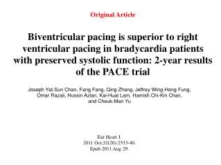
Original Article Biventricular pacing is superior to right ventricular pacing in bradycardia patients with preserved systolic function: 2-year results of the PACE trial Joseph Yat-Sun Chan, Fang Fang, Qing Zhang, Jeffrey Wing-Hong Fung, Omar Razali, Hussin Azlan, Kai-Huat Lam, Hamish Chi-Kin Chan, and Cheuk-Man Yu Eur Heart J. 2011 Oct;32(20):2533-40. Epub 2011 Aug 29.
Biventricular Pacing in Patients with Bradycardia and Normal Ejection Fraction Cheuk-Man Yu, M.D., F.R.C.P., Joseph Yat-Sun Chan, F.H.K.A.M., Qing Zhang, M.M., Ph.D., Razali Omar, M.D., Gabriel Wai-Kwok Yip, M.D., F.A.C.C., AzlanHussin, M.D., Fang Fang, Ph.D., Kai Huat Lam, M.B., B.S., Hamish Chi-Kin Chan, F.R.C.P., and Jeffrey Wing-Hong Fung, M.D., F.R.C.P. N Engl J Med Volume 361(22):2123-2134 November 26, 2009
Introduction • Rright ventricular apical pacing (RVA) can, over time, result in deterioration of left ventricular (LV) function. • Unexpected increased rates of death and heart failure admissions among patients who were randomly assigned DDDR mode in DAVID trial (the Dual Chamber and VVI Implantable Defibrillator) • RVA pacing continues to be practiced because of its easy accessibility and relative stability. Wilkoff BL,et al.2002: JAMA :288:3115-23
Introduction • Preclinical data: Biventricular pacing (BiV) > RVA to preserve myocardial performance. (normal EF) Frias PA et al.2003J Cardiovasc Electrophysiol :14:996-1000. • Acute hemodynamic study: BiV > RVA to preserve LV systolic function (normal EF) Lieberman R et al.2006 J Am Coll Cardiol:48:1634-41. • Clinical study: BiV > RVA to improve exercise capacity & quality of life (LV dysfunction) Kindermann M et al.2006:J Am Coll Cardiol47:1927-37. • Underlying mechanism may be LV systolic dyssynchrony Cojoc A et al. 2006: J Cardiovasc Electrophysiol17:884-9. • BiV pacing -a feasible option in patients with normal LV function as it may attenuate the adverse effect of RVA pacing on LV systolic function.
Hypothesis & Study Design • The Pacing to Avoid Cardiac Enlargement (PACE) study was a prospective, double-blind, randomized, multicenter clinical trial • The study done in four centers at China and Malacia from March 2005 to July 2008. • Atrial-synchronized BiV pacing is superior to RVA pacing in preserving LV systolic function & avoiding adverse LV structural remodeling in patients with standard pacing indication and normal LV ejection fraction
Inclusion criteria • Patients with normal LV ejection fraction (≥45%) who had standard pacing indications • Sinus-node dysfunction • Bradycardia due to advanced AV block
Exclusion criteria • Persistent atrial fibrillation, ACS • PCI or CABG <3 months • Life expectancy of <6 months • Heart transplant recipients • Pregnant women • Patients who fulfilled the eligibility criteria but, implantation of a BiV system was unsuccessful were also excluded
Study Design • Patients received an atrial-synchronized BiV pacemaker capable of delivering RVA pacing or BiV pacing • The RA and RV leads were positioned at the RA appendage and the RV apex and LV lead - posterolateral or lateral branches of the coronary sinus. • Two days after implantation, patients were stratified according to normal or abnormal LV diastolic function • Patients in each group were randomly assigned to receive BiV pacing or RVA pacing, and their pacemakers were programmed accordingly.
Study flowchart Recruitment Randomization Follow up 251 Were screened for pacemaker therapy 89 received BiV pacing (98% BiV pacing) 88 received RVA pacing (97% RVA pacing) 177 underwent randomization 238 Fulfilled the study inclusion criteria 4 died, 3 declined follow up Excluded: inadequate image quality (7), ejection fraction<45% (6) 193 Underwent device implantation 67 Had normal diastolic function 110 Had diastolic dysfunction 81completed 2-yr follow up 82 completed 2-yr follow up (1 -inadequate image quality) 45 declined participation 3died, 3 declined follow up Biv pacing (n=34) RVA pacing (n=55) RVA pacing (n=33) BiV pacing (n=55) 14 patients had a high LV lead pacing threshold (.5 V) and 2 patients had dissection of the coronary sinus without further clinical complications. These 16 patients received conventional dual-chamber pacing and were not included for randomization 2 patients in the BiV pacing group experienced diaphragmatic stimulation and were crossed over to the RVA pacing group at 1 and 7 months, and 1 patient from the RVA group was crossed over to BiV because of heart failure and significant LV dysfunction at 14 month
Study End-points • Primary End-points • LV ejection fraction at 12 months • LV end-systolic volume at 12 months Real-time 3D echocardiography in 90% of the patients, Biplane Simpson’s method in 10% • Secondary End-points • LV end-diastolic volume • 6-min hall walk distance • Quality of life scores (SF-36 health survey questionnaire) For the present 2-year extended follow-up study, all enrolled patients were followed up at 18 and 24 months.
Statistical Analysis • Sample size calculation • Estimated on the basis of the postulated difference in LVEF of 5% between the 2 pacing groups at 12 months (PASS 2000 software,NCSS) • Sample size for the study was 170 patients • Statistical analysis on end-points • Intention-to-treat: patients with ≥3 months follow up were included • Analysis was also performed based on final pacing sites • Two-sided t-test or non-parametric test: for differences in end-points • General Liner Model: potential interaction of clinical factors on primary end-points
Comparison of Primary End-points BiV pacing RVA pacing *P<0.001 vs RVA pacing *P<0.001 vs RVA pacing Absolute difference of EF by 10% Absolute difference of LVESV by 13 ml
Major findings in the study • LV ejection fraction reduced by 13% in the second year of RVA pacing • Eighteen patients in the BiV pacing gp. (20.2%) and 55 in the RVA gp. (62.5%) had a significant reduction of LVEF (of ≥5%, P , 0.001) • Both patients with normal and abnormal baseline LV diastolic function benefited from BiV pacing • No difference in 6-min walk or quality of life between RVA and BiV pacing
Study limitations • Small sample size, not powered at any difference in clinical events • Lower success rate for BiV pacing (92%) than conventional dual chamber pacing • A longer follow-up period is desirable to examine the progressive change in LVESV over time.
Conclusion The PACE study • The first randomized, controlled study showing that In patients with normal systolic function, conventional right ventricular apical pacing resulted in adverse left ventricular remodeling and in a reduction in the left ventricular ejection fraction; these effects were prevented by biventricular pacing.
Original Article Benefit of Early Statin Therapy in Patients With Acute Myocardial Infarction Who Have Extremely Low Low-Density Lipoprotein Cholesterol Ki Hong Lee, Myung Ho Jeong, Ha Mi Kim RN, YoungkeunAhn,Jong Hyun Kim,ShungChullChae,Young Jo Kim,Seung Ho Hur, In WhanSeong,TaekJongHong,DongHoonChoi,Myeong Chan Cho,Chong Jin Kim, KiBaeSeung,Wook Sung Chung,YangSoo Jang, SeungWoonRha,Jang Ho Bae, JeongGwan Cho and Seung Jung Park, J Am CollCardiol 2011;Nov.58:1664–71.
Introduction • Physicians often encounter patients with ACS with LDL-C levels below 70 mg/dl • PROVE IT–TIMI 22 trial revealed no benefit in patients with baseline LDL-C ≤66 mg/dl • Several studies reported that statin therapy resulted in favorable outcomes regardless of baseline LDL-C levels • The influence of baseline LDL-C on the clinical benefit of lipid-lowering therapy remains controversial. • This study investigated whether statin therapy could be beneficial in AMI patients with a baseline LDL-C levels below 70 mg/dl.
Study Design • Analyzed 1,054 patients with AMI who had baseline LDL-C levels below 70 mg/dl and survived at discharge from the Korean Acute MI Registry between Nov. 2005 and Dec. 2007. • They were divided into 2 groups according to the prescribing of statins at discharge (statin gp. n 607; nonstatingp. n 447) • The primary endpoint was the composite of 1-yr. MACE, including death, recurrent MI, target vessel revascularization, and CABG
Clinical outcomes at 6 and 12 Months according to statin medication
Conclusion • Statin therapy in patients with AMI with LDL-C levels below 70 mg/dl was associated with improved clinical outcome • Statin therapy significantly reduced the risk of the composite of the MACE, mainly driven by the risk reduction in cardiac death and coronary revascularization.
Original Article Effects of Hydration in Contrast-Induced Acute Kidney Injury After Primary Angioplasty : A Randomized, Controlled Trial Mauro Maioli, Anna Toso,Mario Leoncini, Carlo Micheletti and Francesco Bellandi Circ CardiovascInterv. 2011;Oct.4:456-462.
Introduction • Intravascular volume expansion is a beneficial measure against contrast-induced acute kidney injury (CI-AKI) in patients undergoing elective angiographic procedures. • Efficacy of this has not yet been established for patients with STEMI, who are at higher risk of CI-AKI after primary PCI. • This study investigated the possible beneficial role of periprocedural intravenous volume expansion in patients with STEMI undergoing primary PCI.
Study Design • Prospective, randomized , 3-arm,single centre study at Misericordia e Dolce Hospital, Prato, Italy. • July 2004 to Dec. 2008, all consecutive patients with STEMI who were candidates for primary PCI • Randomly assigned to 3 groups • preprocedure and postprocedure hydration of sodium bicarbonate (early hydration group) • postprocedure hydration of isotonic saline (late hydration group) • No hydration (control group)
End Points of the Study, Incidence of CI-AKI in High-Risk Patients, and In-Hospital Outcomes inthe 3 Study Groups *Early hydration versus control group, P0.001 (Bonferroni correction). †Early versus late hydration group, P0.015 (Bonferroni correction).
Conclusion • Adequate intravenous volume expansion may prevent CI-AKI in patients undergoing primary PCI. • A regimen of preprocedure and postprocedure hydration therapy with sodium bicarbonate appears to be more efficacious than postprocedure hydration only with isotonic saline.