Download
1 / 66
701 likes | 1.7k Vues
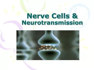
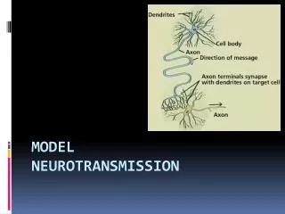
Topic 2d Neurotransmission. Jan 2014. Transmission of Signal Strength at Synapse. Response of postsynaptic cell influenced by amount of neurotransmitter in synapse and number of receptors. Signal Transmission at a Chemical Synapse. Figure 4.16. Neurotransmitters.

E N D
Topic 2dNeurotransmission Jan 2014
Transmission of Signal Strength at Synapse • Response of postsynaptic cell influenced by amount of neurotransmitter in synapse and number of receptors
Signal Transmission at a Chemical Synapse Figure 4.16
Neurotransmitters • More than 50 known substances • Categories • Amino acids • Neuropeptides • Biogenic amines • Acetylcholine • Miscellaneous (gases, purines, etc.) • A single neuron can produce and release more than one neurotransmitter
Dale’s Principles • Candidate NT must be present in the presynaptic terminal • Candidate NT must be released, upon presynaptic stimulus, in amounts sufficient to effect a response in the postsynaptic cell • When added to extracellular fluid, the candidate NT should induce the same changes as the endogenous NT • A mechanism for removal should exist • Effects of drugs on transmission at the synapse must be similar for both endogenous and exogenous NT
Neurotransmitters • Can be excitatory or inhibitory • Most prevalent NT is glutamate • Usually excitatory • GABA (γ-aminobutyric acid) • Usually inhibitory
Agonists/Antagonists http://www.health.bcu.ac.uk/physiology/agonists.gif
Neurotransmitter Action • Inhibitory neurotransmitters • Cause hyperpolarization of membrane • Inhibitory postsynaptic potential (IPSP) • Make postsynaptic cell less likely to generate an AP • Excitatory neurotransmitters • Cause depolarization of membrane • Excitatory postsynaptic potential (EPSP) • Make postsynaptic cell more likely to generate an AP
Glutamate • Used at most fast excitatory synapses in brain and spinal cord • Used at “modifiable” synapses • important in memory and learning
GABA • Used at majority of fast inhibitory synapses in the brain • Many sedatives enhance GABA effects • Glycine has a similar role in the spinal cord
Dopamine • Important in “reward” neural circuits • Dysfunctional in Parkinson’s Disease and schizoprenia
Serotonin • Monoamine NT • ~90% is produced in the intestine; remainder in CNS • Regulates sleep, appetite, memory and learning, etc.
Inactivation of NTs • 4 different mechanisms • Diffusion • Enzymatic cleavage • Uptake by astrocytes • Uptake by presynaptic terminal
Postsynaptic receptors • 2 types • Ionotropic • Directly opens ion channels • Metabotropic • Works indirectly via metabolic changes to the postsynaptic cell to open ion channels
Ionotropic Receptor Function Figure 4.28a
Ionotropic Receptors • Act as ligand-gated ion channels • Some can conduct multiple ions • i.e. Nicotinic Acetylcholine (ACh) receptor • Glutamate AMPA receptor • Equally permeable to K+ and Na+
Fast & Slow Synapses • Until 1980s • Was thought that all synapses were fast • Direct activation of ion channels • Then researchers found: • 2nd messenger mediated cell signalling systems • Slower synaptic actions • norepinephrine
Metabotropic Receptor Figure 4.28b
Receptors for Acetylcholine • Cholinergic receptors • Nicotinic receptor • Ionotropic • Muscarinic receptor • Metabotropic • Linked to ion channel function via G-protein
Receptors for Acetylcholine Figure 4.29
Nicotinic Receptor http://www.nature.com/nrn/journal/v3/n2/images/nrn731-f1.gif
Muscarinic Receptor http://www.ncbi.nlm.nih.gov/books/NBK28014/bin/ch11f10.jpg
Receptors for Acetylcholine Table 4.5
Glutamate Receptors • 3 types • Kainate • AMPA • NMDA
Glutamate AMPA Receptor http://www.chrisparsons.de/Chris/images/AMPA.jpg
Glutamate NMDA Receptor http://www.frca.co.uk/images/NMDA.jpg
Metabotropic Glutamate Receptor http://www.nature.com/embor/journal/v4/n3/images/embor777-f1.gif
EPSPs and IPSPs • Excitatory/Inhibitory Postsynaptic Potentials • Excitatory • Production of a graded depolarization • Brief rising phase and exponential decay • Excitatory synapse usually produces a very small EPSP (<0.5 mV) • Inhibitory • Production of a brief hyperpolarization
EPSPs • Excitatory—depolarization • Na+ and K+ permeability increases at the same time • No action potential • Permeability changes are voltage-independent
IPSPs • GABA • Binds to GABA receptor • Opens Cl- channels • “locks” the membrane at a value more hyperpolarized than the threshold
Second Messenger Systems • G protein coupled receptors (GPCRs) • G protein • Cyclic AMP pathway • DAG & IP3 pathway
http://www.colorado.edu/intphys/Class/IPHY3430-200/image/08-23.jpghttp://www.colorado.edu/intphys/Class/IPHY3430-200/image/08-23.jpg
GPCR structure • Single polypeptide • Main function—activation of G protein • 7 transmembrane domains • Ligands bind near extracellular domain • Several cytoplasmic domains near TM5, TM6, TM7 and maybe TM4 mediate G protein binding • Large protein superfamily
GPCR out 1 2 3 4 5 6 7 In G G protein binding site The G protein binding site is composed of amino acid residues on the C-terminal tail and the intracellular loop between segments 3 and 4, and segments 5 and 6.
G Protein Families http://isoft.postech.ac.kr/Research/POSBIOTM/image/GPCR/table_gpcr1.jpg
http://www.nature.com/scitable/content/ne0000/ne0000/ne0000/ne0000/14707107/U4CP2-2_ActivatedGPCR_ksm.jpghttp://www.nature.com/scitable/content/ne0000/ne0000/ne0000/ne0000/14707107/U4CP2-2_ActivatedGPCR_ksm.jpg
Activated G Protein • Can activate ion channels • Activate 2nd messenger cascades • Cyclic AMP • Phospholipase C pathway
Inactivation of G protein • α subunit of G protein contains a GTPase • GTP degraded to GDP • α subunit reassociates with β and γ subunits
Activation of muscarinic receptors • Cholinergic synapses in cardiac tissue • GPCR activates G protein • α subunit + GTP dissociates • β and γ subunits diffuse through membrane to activate K+ channels—causing IPSPs
Cyclic AMP cascade • G protein activates adenylyl cyclase • Catalyzes conversion of ATP to cyclic AMP • Cyclic AMP activates protein kinases • Protein kinases phosphorylate other proteins • Activation of ion channels • Metabolic changes • Changes in transcription factors http://t3.gstatic.com/images?q=tbn:ANd9GcQev_GY0IC6y5BsuY4GoQ-8UaWogpktHlZpStl29YimNMDMXGP7
http://employees.csbsju.edu/hjakubowski/classes/ch331/signaltrans/camp.gifhttp://employees.csbsju.edu/hjakubowski/classes/ch331/signaltrans/camp.gif
Cyclic AMP http://psychology.jrank.org/article_images/psychology.jrank.org/neurotransmitters-and-neuromodulators.9.jpg